Introduction
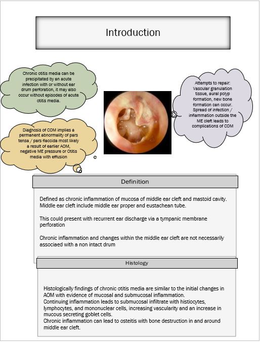
Chronic otitis media is defined as chronic infection of mucosa lining the middle ear cleft and mastoid cavity. Middle ear cleft includes the middle ear proper and the eustachean tube. Diagnosis of Chronic otitis media implies a permanent abnormality of pars tensa / pars flaccida portion of the ear drum.
This condition most likely is a result of:
1. Earler episodes of acute otitis media
2. Negative middle ear pressure
3. Otitis media with effusion
It should be stressed that chronic inflammation of middle ear cavity is not necessarily associated with a perforated ear drum.
Complications of COM is mostly due to inflammation extending outside the confines of the middle ear cleft.
The following features are some of the evidence of reparative process:
Vascular granulation tissue
New bone formation
Use of microscope is performing routine otological examination is common these days. It allows both hands of the surgeon free to clean the external canal, suck out the secretions and to examine the ear drum under magnification. This has also done away the concept of tubotympanic and atticoantral disease. The tern safe and unsafe ear is also not used these days.
Initially tubotympanic middle ear disease which involves middle ear cleft alone is considered to be a safe disease as dangerous complications were not common where as attico antral disease that involves the attic region of the middle ear cavity is considered tobe a dangerous variety of otitis media because rates of complications were found tobe a bit high.
High incidence of complications in atticoantral disease is due to:
1. The proximity of the area to skull base
2. Crowding of structures seen in this area
But current understanding of pathophysiology of chronic otitis media after the advent of routine microscopic examination of the ear has undergone lots of changes that has been incorporated into the currently acceptable classification of chronic otitis media types.
