Diagnosis and Assessment
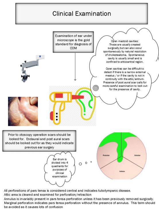

Examination of ear under microscope is the gold standard in the diagnosis of COM. Microscope allows the examiner to manipulate the ear, remove wax, clean up the secretions as both the hands are free. Ear drum can be visualized under magnification.
Otoscopy performed by using a microscope and appropriate aural speculum faciliates accurate diagnosis of COM. After a good aural toilet, the clinician can assess all areas of external ear and the ear drum.
Operation scars:
Prior to performing otoscopy the presence of operation scar should be looked out for in the post aural region / end aural region. This will help in accurately assessing the mastoid cavity if canal wall down surgery has been performed.
Mastoid cavities:
Cavities in the mastoid area could be created surgically or by the presence of cholesteatoma. Open mastoid cavities could be difficult to detect if the external canal is narrow or if the cavity is not continuous with the attic / antrum.
Site of the pathology:
This can be accurately described by visualizing the area of ear drum involved. In order to provide accurate description of the area of ear drum involved, the pars tensa is divided into 4 quadrants. They include:
Antero inferior
Postero inferior
Antero superior
Postero superior
Perforation of ear drum can be accurately described by describing the quadrants involved in the perforation.
