Why myringoplasty fails?
Contents
Synonyms:
Myringoplasty failure , Type I tympanoplasty failure
Definition:
Myringoplasty is defined as the procedure where in an autologous graft material is used to patch a perforated ear drum. There are two accepted techniques involved in this procedure:
1. Overlay technique (On lay)
2. Underlay technique (Under lay)
Both these techniques have been followed for quite some years with consistent results.
Overlay technique:
is otherwise known as lateral graft tympanoplasty. In this technique skin is used as the grafting material. This procedure requires complete removal of all squamous epithelium from the entire outer surface of the drum head, the entire annular ligament, and the most inaccessible anterior sulcus. Skin is used to refashion neotympanum.
Underlay technique:
is otherwise known as medial graft tympanoplasty. This is the commonest type of myringoplasty performed. Temporalis fascia is used as the graft material, and is placed under the tympanomeatal flap which has been elevated. The tympanomeatal flap is then repositioned. The graft material lies medial to the ear drum. It is also tucked under the handle of malleus.
Common causes of myringoplasty failures include:
1. Upper air way infections
2. Type of surgical procedure
3. Type of tissue used to graft the perforation
4. Trapped epithelial seed cells post operatively
5. Overlay graft technique
Exposure of ear drum:
Adequate exposure of ear drum is a must before proceeding with surgery. Even though it is not necessary to see all areas of drum head in one view, different areas of drum should be seen by simple manipulation of position of the patient / microscope etc. In 5 - 10 % of patients there may be a prominent bulge in the anterior canal wall obscuring the anterior rim of the ear drum and the anterior portion of the annulus. Myringoplasty performed under these conditions may fail because the graft could medialize in the anterior recess area, which is a really deep area. Medialisation of graft is common in these conditions. This scenario can be prevented in these patients by elevation of Wright Guilford flap in these patients. This flap is raised from over the bulge of the anterior canal wall through an incision made circumferentially using a Rosen's knife about 7mm lateral to the ear drum. The skin and periosteum are elevated from the bony hump. In majority of these patients this procedure alone brings the anterior margin of the ear drum and anterior annulus tympanicus in to view. If still these structures are not visible then the bony hump can be shaved using a diamond burr.
Preparation of drum head before grafting:
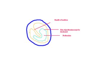
For a successful underlay technique the drum head must be properly prepared for receiving the graft material (temporalis fascia in this case). This involves freshening the edges of the perforation. In fact a rim of ear drum tissue is removed around the perforation. The handle of malleus is identified, and if possible is freshened at this stage. If the perforation is small then this step will have to wait till the tympanomeatal flap is elevated. The under surface of the tympanic membrane must be scraped using a instrument called drum scraper. The aim is to create raw area on the undersurface of the ear drum facilitating a better graft take.
Presence of secondary pathology in the middle ear:
The following disorders of the middle ear can lead to graft rejection:
1. Tubal obstruction
2. Presence of cholesteatoma
3. Presence of tympanosclerosis
4. Presence of adhesions binding the handle of malleus to the promontory
5. Ossicular chain necrosis
Positioning of graft:
In underlay technique of myringoplasty the graft must be positioned in such a way that it lies under the handle of malleus. The handle of malleus is exteriorised. This method prevents lateralisation of the graft due to pull by the migrating squamous epithelium. If it is inserted medial to the handle of the malleus this complication can be prevented.
The skin lining of the external canal must be preserved for better healing of the perforation. The attic area must be explored to rule out attic pathology like cholesteatoma.
The handle of malleus must be denuded off its mucosal covering, and it should be freed from adhesion if any with the promontory. This will increase the chances of graft take. Gelfoam packs must be placed in the middle ear cavity in order to enhance the nutritional status of the graft material, it also helps to prevent medialisation of the graft.


