Tympanoplasty procedure
Contents
- 1 Definition:
- 2 Canalplasty:
- 3 Meatoplasty:
- 4 Ossiculoplasty:
- 5 Tubal function tests
- 6 Radiological investigation:
- 7 Pre op preparation:
- 8 Anesthesia:
- 9 Surgical approach:
- 10 Endaural approach:
- 11 Post aural approach:
- 12 Selection of approach:
- 13 Grafting techniques:
- 14 Total overlay:
- 15 Transcanal myringoplasty:
- 16 Endaural approach:
- 17 Post aural approach:
Definition:
Tympanoplasty is defined as the surgical procedure which enables reconstruction of middle ear cavity and the conducting ossicular system (tympano- ossicular system).
The following surgical procedures are components of tympanoplasty:
1. Canal plasty
2. Meatoplasty
3. Myringoplasty
4. Ossiculoplasty
Canalplasty:
Is widening of the external auditory canal. It should be considered to be an integral part of myringoplasty. This procedure should be performed before grafting anterior perforations of tympanic membrane as it gives necessary surgical access for repair. This procedure facilitates healing, cleansing and if needed a second stage ossiculoplasty.
Meatoplasty:
Is used to enlarge the opening of lateral cartilagenous portion of external auditory canal. This enlargement should be in proportion to the size of the medial bony external auditory canal.
Ossiculoplasty:
This procedure is used to reconstruct the damaged middle ear sound conducting ossicular system.
Aims of this surgical procedure:
a. Disease eradication
b. Restoration of middle ear aeration
c. Reconstruction of sound transmission mechanism
d. Creation of self cleansing dry cavity
Desired Preoperative investigations:
Tubal function tests
Tubal funtion tests is very important for proper surgical planning and to assess the chance of a possible hearing improvement. If tubal function tests are negative, blockage if any at the tympanic end of the eustachean tube. If any scar tissue is present in that area it should be meticulously removed to pave way for better ventilation of the middle ear cavity.
Radiological investigation:
Xray of temporal bones / CT scan temporal bones will be a reliable indicator for pneumatization of temporal bone. A well pneumatized temporal bone indicates a normally functioning eustachean tube. Sclerosed mastoid indicates supressed due to infantile otitis media or blocked eustachean tube.
Temporary closure of perforation with wet gelfoam: This helps to assess the condition of ossicular chain / status of round and oval windows. If hearing improves when the perforation is temporarily occluded with a gelfoam plug then it indicates normally functioning middle ear sound conduction mechanism.
Fistula test:
Should always be performed before surgery. If it is positive then surgery should be deferred as this would lead to dead ear.
Pre op preparation:
1. Surgery should be ideally performed in a dry ear. The ear must be cleaned before surgery. Systemic / topical antibiotics should be administered to reduce the likelihood of infection.
2. A persistently draining ear is a problem. If the secretion is purulent then it should be swabbed and sent for culture and sensitivity. This will help in prescription of drugs to which the microbes are sensitive to. A predominantly clear mucoid secretion from the ear is definitely related to hyperplastic changes of middle ear mucosa, hence ear swab for culture sensitivity is not necessary.
3. Hair is shaved above and behind the ear about 2 cms. The ear canal is plugged with sterile cotton when the area of surgery is disinfected with povidone iodine. The external auditory canal is not disinfected.
Anesthesia:
Local / General
Position: Supine. The head should be positioned slightly lower than the plane of the table. The angle between the head and shoulder should be between 100 - 130 degrees to allow adequate working space for the surgeon's hands.
Surgical approach:
Transcanal approach:
In this approach surgery is performed through the ear speculum inserted into the external auditory canal. This approach is indicated in:
a. Patients with wide external auditory canal
b. There is no overhanging bony wall obscuring the rim of the perforation
Endaural approach:
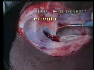
In this approach the incision is made betweenthe tragus and helix. An endaural speculum is introduced to widen the entrance into the external auditory canal. If posterior overhang is present it can be easily burred out. If this approach is used better anterior visualization of the ear drum is possible.
Post aural approach:
Mastoid bone and the external auditory canal is approached via a post aural incision of William Wilde.
This approach is useful in cases of:
1. Narrow external auditory canal as it would improve exposure and facilitate canalplasty
2. In perforations involving the anterior portion of the ear drum
Selection of approach:
Transcanal approach: is mostly used for repairing large acute traumatic perforations. The external canal should be fairly wide, and the rims of the perforation should be clearly visible.
End aural approach:
Perforations involving the posterior portion of ear drum can be adequately visualized by endaural approach.
Postaural approach: Is reserved for those perforations whose margins could not be entirely visualised through intact external canal.
Grafting techniques:
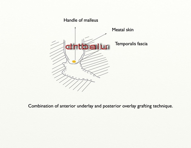
Two types of grafting techniques are available i.e. overlay and underlay. Overlay: The term overlay means that the graft has been placed over the bony tympanic sulcus, or over a bony ledge carved newly for this purpose when sulcus is absent. The overlaid fascia is supported by the presence of new / old annulus and is held in position by a remnant of tympanic membrane if still present. Underlay: The term underlay indicates the position of the graft under the tympanic membrane and surrounding bone. Combination of anterior underlay and posterior overlay:For this type of grafting procedure to succeed, there should be an anterior remnant of the tympanic membrane (atleast of the fibrous annulus). Anteriorly the graft is placed under the anterior remnant of the tympanic membrane and under the lateral wall of protympanum. Posteriorly, it is placed over the posterior tympanic sulcus and under the remnant of the tympanic membrane. With the exception of perforations limited to the anteroinferior quadrant, the graft lies under the malleus handle.
Total overlay:
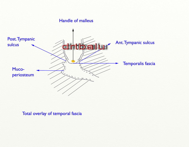
This technique is useful when there is no remnant of the tympanic membrane is present. A new bony sulcus is drilled to support the fascia at the lateral opening of the tympanic cavity. The graft rests over the sulcus and underneath the malleus handle. The edges of the graft are covered along the canal wall by meatal skin.
Transcanal myringoplasty:
Indication:
1. Used to repair large acute traumatic perforations of ear drum
2. Used to repair dry central perforations.
Anesthesia: Ideally local
Ear speculum should be used to straighten the external auditory canal.
Perforated margins are outfolded.
Repositioned perforated margins are held in position by intra and extratympanic fixation using gelfoam.
Procedure:
1. Perforation is visualized using aural speculum. Use of aural speculum straightens the external canal.
2. The speculum is fixed by holding it in the left hand
3. Outfolding of perforated margins: The edges of the perforation are outfolded using a 1.5mm, 90 degree hook.
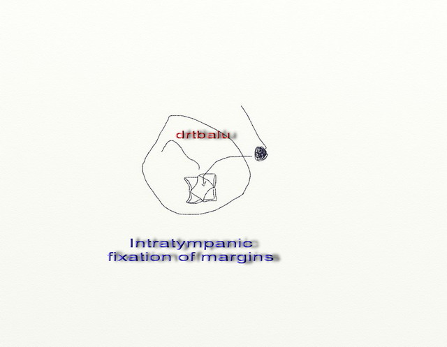
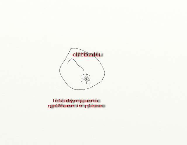
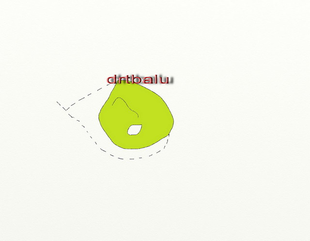
4. Intratympanic fixation of perforation margins: Outfolded perforation margins are kept in position by intratympanic gelfoam pledgets soaked in Ringer's solution.
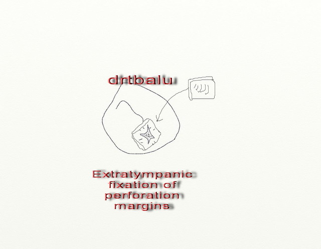
5. Extratympanic fixation of perforation margins: A piece of gelfoam is wet with Ringer lactate solution is used to stabilize the outer surface of lacerated tympanic membrane.
Endaural approach:
This technique is used for repairing posterior perforations, whose margins can be clearly seen through external auditory canal.
Anesthesia: Local
Incision: Endaural Lempert's incision
The incision is 5 - 10 mm long and is performed between the tragus and crus of helix.
Tympanomeatal flap elevation and freshening of perforation margins: The skin incision for tympanomeatal flap is carried from the tympanic annulus at 7 'o clock and 12 'o clock in an ascending spiral fashion to meet the endaural incision. The anterior limb of the incision extends just anterior to the lateral process of the malleus. The edges of the perforation is freshened before elevation of the flap.
After elevation of tympanomeatal flap, gelfoam pledgets soaked in Ringer's lactate is placed in the hypotympanum. Mobility of ossicular chain is checked.
Graft is inserted beneath the perforated drum and above the posterior tympanic sulcus.
Post aural approach:
Indication: This approach is mainly used for anterior perforations of ear drum whose margins are obscured by bony overhang. In most of these patients canal plasty need to be performed.
Anesthesia:
General anesthesia.
Incision:
Post aural skin incision of William wild is used. It is carried out along the hairline and behind the mastoid tip. The incision is initially made only skin deep preserving underlying fascia and periosteum.
Elevation of periosteal flap: Periosteal flap is then elevated from the bone using a periosteal elevator.
Exposure of external auditory canal:
While the periosteal flap is being elevated, the external auditory canal can be entered.
If proper visualization could not be achieved canalplasty needs tobe performed.
Canalplasty: In this procedure the external bony canal is enlarged using diamond burrs, eliminating all bony overhangs. Care should be taken not to breach the temporomandibular joint situated anteriorly.
The rim of the perforation is freshened. Graft inserted, using underlay technique. Flap repositioned.


