Tumors of pinna and external auditory canal
Introduction:
Tumors of external ear could arise from the pinna or external auditory canal. Tumors can be benign or malignant. Among carcinomas involving the external ear 85% involve the pinna while 10% involve the external auditory canal.
Benign tumors:
Preauricular sinus / cyst:
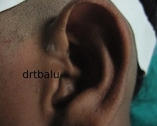
This is caused by faulty union of hillocks of the first and second branchial arches during development of pinna. Preauricular sinus usually presents as a small opening in front of the crus of helix. It has a branching tract that is lined by squamous epithelium. When this tract gets blocked it leads to the formation of retention cysts. This cyst could ultimately be infected giving rise to pain and increase in the size of swelling. Surgery is usually indicated if there is swelling and infection. The sinus / cyst should be completely excised to avoid recurrence.
Simple preauricular cysts should not be confused with that of first branchial cleft cysts. Branchial cleft anomalies are very closely associated with the external auditory canal, ear drum, angle of mandible and facial nerve. Misdiagnosing a first branchial abnormality for simple sinus tract / cyst could cause the unsuspecting physician at risk for damagig the facial nerve.
Patients with preauricular pits / cysts must be examined for the presence of other congenital abnormalities.
Preauricular skin tages:
These are epithelial mounds / pedunculated skin tags that rise in front of the pinna around the tragus. These structures dont have bony, cartilaginous or cystic components. It also does not communicate with the external auditory canal / middle ear.
Investigations:
In the presence of abscess Incision and drainage should be performed and pus should be sent for culture and sensitivity.
Imaging:
Imaging is not indicated for routine preauricular cysts and sinuses. It is indicated in patients who present with pits / fistulas in atypical regions, those with cartilage duplication around the external canal that extends into the parotid, and in those patients with recurrent parotid swelling.
Patients with preauricular cysts / pits / branchial cleft cyst should undergo renal ultrasonography to rule out branchio-oto-renal syndrome.
Syndromes associated with preauricular pits / cysts / sinuses / tags:
1. Branchiootorenal syndrome - perauricular sinus
2. Beckwith-Wiedemann syndrome - Preauricular sinus with asymmetric earlobes
3. Mandibulofacial dysostosis - Auricular pits / fistulas
4. Oculoauriculovertebral dysplasia - Preauricular tags
5. Chromosome arm 11 q duplication syndrome - Preauricular tags or pits
6. Chromosome arm 4 p deletion syndrome - Preauricular dimples or skin tags
7. Chromosome arm 5 p deletion syndrome - Preauricular tags
Management:
Antibiotics - Cephalexin, amoxycillin and clavulanate potassium are indicated in patients with cellulitits from infected preauricular pits.
Incision and drainage may be required in patients with abscess formation. Staphylococcus aureus is the most common bacteria isolated in these infections. Proteus, streptococcus and peptococcus have also been isolated. Needle aspiration can be performed as an alternative to incision and drainage in the presence of abscess.
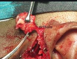
Complete excision of sinus tract should be performed if infection is persistent / recurrent. Recurrence can be avoided by ensuring complete resection of the sinus and tract along with associated perichondrium. Dissection should be performed up to the level of temporalis fascia to ensure complete resection.
Sebaceous cyst:
The common site for sebaceous cyst close to pinna is the post auricular sulcus, or below and behind the ear lobule. This is also known as epidermal cyst, epithelial cyst, keratin cyst or epidermal cyst. This condition is caused by implantation of epidermal elements with subsequent cystic transformation. Epidermoid cyst present in the retroauricular region should be differentiated from that of lipoma and hemangioma.
Hemangioma:
These are congenital tumors seen in the post auricular region. Hemangiomas are of two types:
Capillary hemangiomas - This is a mass of capillary sized blood vessels and may present as a port wine stain. It does not regress spontaneously.
Cavernous hemangioma - This is also known as straw berry tumor. It is made up of endothelial line spaces filled with blood. It increases rapidly in size during the first year of life and regresses there after. it could fully disappear by the age of 5.
Dermoid:
This presents as cystic mass, rounded over the upper part of mastoid behind the pinna. Histologically dermoid cyst can be classified as:
Dermoid cyst
Epidermoid cyst
Teratoma
This is a benign congenital tumor formed by cells abnormally separated at the period of fusion of tissues. The cyst is encapsulated containing stratified epithelium lined by laminated keratin material containing adnexial structures of skin such as sebaceous gland, sweat gland, hair follicles and hair.
These cysts can be managed by complete excision.
Keloid:
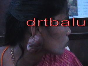
This usually develops following trauma like piercing the ear lobule. This condition shows genetic susceptibility. It is common among blakc races. It usually presents as a pedunculated tumor.
It can be treated by surgical excision. Steroid injection into the surgical site can also be attempted.
Papilloma:
Also known as wart. It usually presents as a tufted growth of a flat grey plaque. It is rough on touch. It is caused by viral infections. Can be managed by surgical excision or by cautery.
Cutaneous horn:
This is a type of papilloma which involves heaping up of keratin and presents as a horn shaped tumor. It is seen at the rim of antihelix in elderly people. Can be treated by surgical exicision.
Keratoacanthoma:
It is a benign tumor which clinically resembles a malignant one. It appears as a raised nodule with a crater in the centre. Treatment is by excision biopsy.
Neurofibroma:
This usually presents as a non tender, firm mass. May also be associated with von Recklinghausen disease. Treatment is usually by surgical excision.
Malignant lesions involving pinna include:
Squamous cell carcinoma:
Usually helix is involved. It presents as a painless nodule or ulcer with everted edges and basal induration. Regional nodal metastasis occur rather late. It is more common in males. Wide excision followed by irradiation is the treatment modality.
Basal cell carcinoma:
Common site of involvement are the helix and the tragus. This tumor is most common in men beyond the age of 50. It presents usually as a painless nodule with a central crust. Removal of this crust results in bleeding. Ulcers usually have a raised edge. Ulcers could penetrate deeper involving cartilage / bone. Nodal metastasis dont occur.
Superficial lesions not involving cartilage can be irradiated thereby avoiding cosmetic deformity. Lesions involving cartilage would require surgical excision.
Melanoma:
This malignant tumor can arise anywhere over the auricle. This lesion is common in men with light complexion and who are exposed to sun. Nodal metastasis is seen in nearly half of these patients.
Superficial melanoma less than 1 cm in diameter if situated over the helix can be managed by wedge resection and primary closure. Melanoma larger than 1 cm and recurrent melanomas can be treated by resection of pinna, parotidectomy and radical neck dissection.
Tumors of external auditory canal:
Benign tumors:
Osteoma:
This tumor arises from cancellous bone of the external canal. It appears as a single smooth bony hard pedunculated tumor. This mass usually arise from the posterior wall of the osseous meatus near its lateral end. Treatment is surgical removal using electric drill.
Exostosis:
These are multiple and bilateral bony lesions. These lesions present as smooth sessile bony swellings in the deeper portion of tbe external auditory canal close to the ear drum. These lesions arise from the compact bone. These lesions are commonly seen in those individuals exposed to entry of cold water (divers / swimmers). Males are affected three times more commonly than females.
Asymptomatic cases need not be treated. Larger lesions that impair hearing need to be excised. These lesions could lie close to the facial nerve and hence this fact should be taken into account during surgery for removing these masses.
Ceruminoma:
These are tumors of modified sweat glands which secrete cerumen. These lesions present as smooth, firm, skin covered polypoidal swelling in the outer portion of the meatus. It is generally attached to the posterior or inferior meatal wall. It cause obstruction of the external canal leading on to retention of wax and other debris. Malignant version of these tumors out number their benign counterpart by a ratio of 2:1.
Since this tumor has a tendency to recur, wide surgical resection of the lesion is preferred. If there is suspicion of malignancy post op radiotherapy needs to be administered.
Malignant tumors involving the external canal:
Squamous cell carcinoma;
This is seen in cases of long standing ear discharge. This fact could be controversial and should in no way construed that CSOM is a premalignant condition. It can be a primary lesion arising from the skin lining of the external canal or a secondary lesion arising from middle ear cavity tumor extension.
These lesions usually present as blood stained discharge. Clinical examination reveals that external canal appears ulcerated with the presence of bleeding polypoidal mass / granulation tissue. Facial nerve paralysis is also common in these patients. Regional nodal metastasis is also common.
Basal cell carcinoma:
These lesions arise from the external canal. Clinical picture is more or less similar to that of the squamous variety. Biopsy of the lesion is diagnostic.
Wide surgical excision is the preferred treatment modality.


