Temporal bone fractures
Contents
Introduction:
Fractures involving temporal bones are seen in nearly 20% of all head injuries. Commonly temporal bone fractures are unilateral. Since fractures of this bone is associated with other sinister injuries, it has been given least importance. From the functional stand point, early involvement of otolaryngologist in these patients will reduce the incidence of long term sequel.
Common sequel of temporal bone fractures:
1. Facial nerve palsy
2. Damage to cochleo vestibular apparatus causing sensorineural hearing loss
3. Conductive hearing loss due to ossicular disruption
4. Balance disturbance
5. Tinnitus / vertigo
6. C.S.F. leak
7. Perilymph fistula
8. Post traumatic endolymphatic hydrops
9. Cholesteatoma
10. Menigocele / encephalocele
11. Otogenic meningitis
12. Injuries to cranial nerves like VI, IX and cranial nerves up to XI
13 Vascular injuries i.e. injuries to internal carotid artery and sigmoid sinus
Classification:
Ulrich's classification:
Ulrich in 1926 classified temporal bone fractures into either transverse or longitudinal. Since many of the clinically observed features could not be simply explained by these fracture types terms like oblique and mixed types have been added.
Longitudinal fractures:
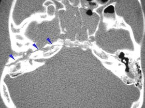
This is the commonest type accounting for 80% of all temporal bone fractures. These fractures are caused by lateral blows like temporal or parietal types.
The fracture line parallels the long axis of the petrous pyramid. It starts from the squamous portion of the temporal bone, extends through the postero superior portion of the external auditory canal, continues across the roof of the middle ear space, anterior to labyrinth to end anteromedially in the middle cranial fossa close to foramen lacerum and ovale.
Signs / Symptoms:
1. Bleeding from external canal due to laceration of skin and ear drum
2. Haemotympanum (conductive deafness)
3. Fractures involving the bony portion of external canal
4. Ossicular chain disruption causing conductive deafness.
5. Facial palsy (rare) 20% usually at the level of horizontal segment distal to geniculate ganglion
6. CSF otorrhoea (usually temporary)
7. Sensorineural hearing loss can occur due to consussion
Transverse fractures:
This type of fracture comprises about 20% of all temporal bone fractures. They are usually caused by frontal / parietal blow. Rarely it could result from occipital blow also. The fracture line runs at right angle to the long axis of the petrous pyramid. Usually it starts in the middle cranial fossa close to foramen lacerum, it crosses the petrous pyramid transeversely to end at the foramen magnum. Sometimes it may run across the internal acoustic meatus causing damage to auditory and facial nerves.
Clinical features:
1. Sensorineural hearing loss due to damage to 8th cranial nerve
2. Facial palsy due to damage of facial nerve
3. Vertigo
4. Labyrinthitis ossificans (this should be borne in mind before performing cochlear implant in these pts)
Another classification system has been devised by Kelly and Tamito facilitate better clinical correlation of fracture geometry with clinical outcomes. In this type of classification fractures of temporal bone has been subdivided into otic capsule violating type and otic capsule sparing types. CSF leak and sensori neural hearing loss are common in otic capsule violating fractures.
Mechanical factors:
1. The human skull deforms before actual fracture occurs
2. The mechanical tensile strength of skull do not deteriorate with age
3. Temporo parietal region requires as much force as frontal region for fracture to occur
Work up:
Imaging:
HRCT: HRCT of temporal bone is useful in assessing injuries complicated with CSF leak, facial palsy or suspected vascular injury. Usually 1mm cuts in both axial and cornal planes must be performed. Bone window cuts would be really useful.
It is also indicated when surgical intervention for otologic complications following temporal bone fracture becomes necessary.
It is indicated in patients with persistent cranial nerve injuries following skull base fracture.
CT angiography:
This is indicated in evaluation of petrous carotid injury.
MRI:
Helps in identification of intralabyrinthine haemorrhage, brain stem injury and nerve compression.
General clinical presentation of temporal bone fractures:
Neurologic injuries: Since tremendous forces are required to cause fractures of temporal bone, these patients invariably present with multiple injuries like associated non temporal skull fractures and injuries to head and neck.
Commonly seen neurologic injuries in these patients are:
1. Subdural hematoma
2. Subarachnoid hemorrhage
3. Contusion of brain
4. Tension penumocephalus
5. Injury to facial nerve
6. Injury to vestibulocochlear nerve
Facial palsy: is commonly seen in transverse fractures of temporal bones. Injury to facial nerve is rather uncommon in pediatric age group patients. This has been attributed to the increased flexibility of pediatric skull.
The timing of onset of facial palsy has an important bearing on its outcome. Immediate palsy invariably indicates total transection of the nerve, while delayed palsy indicates the development of neuronal oedema / hematoma with nerve compression inside a non expanding facial nerve bony canal. In any case it is really worthwhile to open up the mastoid in all cases of traumatic facial nerve palsy in order to have a look.
CSF leak: is mostly in the form of otorrhoea. In the acute phase it could be missed as it would be admixed with blood. In patients with intact ear drum the csf would be shunted out via the eustachean tube and hence could easily be missed. Hence a high degree of suspicion is important in diagnosing these patients.
Following signs could point towards the diagnosis:
1. Halo sign
2. Reservoir sign
3. Presence of Beta - 2 - transferrin in the fluid is diagnostic
CSF leak is three / four times more common in patients with otic capsule violating type fractures.
Vascular injuries: All temporal bone fractures involving the jugular fossa / sigmoid groove always resulted in breech of internal jugular vein wall. Traumatic dissecting aneurysm of internal carotid artery can also be seen in patients with otic capsule violating injuries.
Delayed sequel:
Menigocele / encephalocele: are late manifestations of temporal bone injuries. It can manifest as late onset CSF otorrhoea, unilateral clear middle ear effusion, or recurrent meningitis. The delay can range from 1 - 20 years. These delayed herniations can be approached in the same manner as acute ones. Defects must be sealed after reduction of meningocele / encephalocele.
Cholesteatoma: is one of the late complications of temporal bone fractures. This could be due to traumatic implantation of epithelial elements during injury into the middle ear cavity.


