Surgical Anatomy of Thyroid Gland
Surgical anatomy of thyroid:
The thyroid gland has two lobes the right and the left. These lobes are connected in the midline by a sleeve of thyroid tissue known as the isthmus. The whole gland is covered anteriorly by infrahyoid group of muscles.
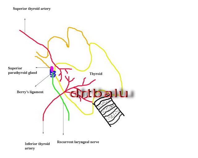
Blood supply of thyroid: Major blood supply to thyroid gland arises from the superior thyroid artery a branch of the external carotid artery, and inferior thyroid artery by way of the thyrocervical trunk. Venous supply accompanies the arteries. A middle thyroid vein directly drains into the internal jugular vein.
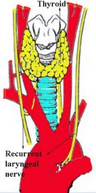
Recurrent laryngeal nerves and their relationship to the thyroid gland: The recurrent laryngeal nerve innervate the intrinsic muscles of larynx. It also provides sensory innervation to the glottis. The recurrent laryngeal nerve arises from the vagus at the level of subclavian artery on the right side and at the level of the aortic arch on the left. The nerves then turn superio medially and runs towards the tracheo oesophageal groove. As the recurrent laryngeal nerve ascends the tracheo oesophageal groove it is intimately related to the inferior thyroid artery. The nerves may pass superficial or deep between the branches of the inferior thyroid artery.
The recurrent laryngeal nerve as it travels in the tracheo oesophageal groove, it comes into intimate contact with the posterior portion of the thyroid gland. It is always better to identify the nerve at the level of cricothryoid joint, at which point it enters the larynx. Injury to this nerve should be prevented during surgery at all costs, as this will cause vocal cord paralysis. Damage to recurrent laryngeal nerves on both sides will cause stridor necessitating tracheostomy due to bilateral abductor palsy. Non recurrent laryngeal nerve: arises directly from the cervical portion of the vagus at about the level of the larynx and enters it at the level of the cricopharyngeal joint. Majority of these nerves occur on the right side and is commonly associated with an anomalous retro esophageal subclavian artery. Effects of Berry's ligament on the course of recurrent laryngeal nerve: The pretracheal fascia that covers the thyroid gland condenses and attaches the thyroid gland to the upper two tracheal rings is known as the Berry's ligament. The recurrent laryngeal nerve often passes through this layer to enter the larynx.
Superior laryngeal nerve: arise from the inferior vagal ganglion (nodose) and descend inferiorly deep to the carotid system. As the superior laryngeal nerve descends towards the thyrohyoid membrane they pass anterior to the cervical sympathetic trunk and posterior to the carotid system. Friedman proposed a classification to account for the anatomic variations of superior laryngeal nerve.
They are:
Type I: The nerve runs superficial to the inferior constrictor muscle.
Type II: The nerve penetrates the lower part of the inferior constrictor muscle.
Type III: The nerve penetrates the superior part of the inferior constrictor muscle.
The superior laryngeal nerve travels in close proximity to the superior thyroid artery. This nerve should be protected by the surgeon at all costs.
Injury to this nerve will cause minor degrees of voice change since this nerve supply the cricothyroid muscle. It patient will not be able to raise the pitch of his voice. This becomes really troublesome for a singer. It also supplies sensory innervation to larynx.
Parathyroid glands:
During surgery every effort should be made to identify and preserve the parathyroid glands. These glands are 4 in number. The superior parathyroids embryologically arise from the 4th pouch, while the inferior parathyroids arise from the 3rd pouch. The superior parathyroid glands lies near the cricothryoid joint, at the intersection between the recurrent laryngeal nerve and the inferior thyroid artery. The inferior parathyroids are variable in position because it has to migrate long distances due to the position of the thymus gland. Commonly they are located close to the inferior thyroid pole. The parathyroid glands are supplied by branches from the inferior thyroid artery, hence it should be protected.

