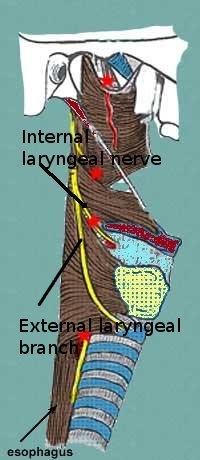
Superior laryngeal nerve
This branch from the vagus supplies the main sensory innervation of larynx in the glottic and supraglottic regions with some minimum contribution to posterior subglottic area. It branches out of the vagus nerve just below the nodose ganglion. It is branched from the medial aspect of the vagus nerve. It lies superficial to the superior cervical ganglion form which it receives sympathetic supply. This nerve also supplies the carotid body. Its motor innervation supplies the cricothyroid muscle. Traditional belief is that paralysis of superior laryngeal nerve causes bowing and flaccidity of true vocal cords with decreased vocal range and laryngeal rotation. According to Sulica this premise is not entirely accurate. In human larynx the superior laryngeal nerve has complex functional inter relationship with the recurrent laryngeal nerve. Hence the degree of compensation of these two nerves following injury is not entirely straight forward.
The average length of superior laryngeal nerve is about 2 cm in males and 1.5 cms in females. It divides into an internal and external laryngeal branches.
Internal laryngeal nerve branch:
is predominantly sensory in nature. This nerve runs parallel and medial to the superior laryngeal artery. At the level of greater cornu of hyoid bone it turns medially, passing deep to thyrohyoid muscle. This nerve enters the larynx through the thryohyoid membrane just above the superior border of inferior pharyngeal constrictor muscle. After entering into the larynx this nerve divides into three branches i.e. superior, middle and inferior. The superior division divides into two / three branches supplying sensations to the lingual surface of epiglottis, lateral aspect of glosso epiglottic fold. The middle division innervates the aryepiglottic fold, vocal folds, vestibular folds and the posterior aspect of arytenoid. The inferior division is the largest of the branches of superior laryngeal nerve. It lies along the medial aspect of pyriform fossa. It is this nerve which is blocked when pyriform fossa block is given for endolaryngeal surgical procedures. This branch supplies the interarytenoid muscle. This nerve gives out a branch that communicates with the recurrent laryngeal nerve (Galen's loop).
External branch of superior laryngeal nerve is smaller in caliber when compared to the internal branch. It supplies motor fibers to the cricothyroid muscle. It may provide occasional supply to the thyroarytenoid muscle. Rarely it may also provide sensation to the glottis. This nerve arises from the superior laryngeal nerve at the level of greater cornu of hyoid bone. At this level it lies just posterior to the superior thyroid artery.
Kierner classified the superior laryngeal nerve into 4 types depending on the relationship of its external branch to the superior pole of thyroid gland.
Type I nerve: In this type the external branch of superior laryngeal nerve cross the superior thyroid artery about 1cm above the superior pole of thyroid gland.
Type II nerve: In this type the external branch of superior laryngeal nerve crosses the superior thyroid artery within 1 cm of the superior pole of thyroid gland.
Type III nerve: In this type the external branch of superior laryngeal nerve crosses the superior thyroid artery under cover of the superior pole of thyroid gland.
Type IV nerve: In this type the external branch of superior laryngeal nerve descends dorsal to the superior thyroid artery and crosses its branches just superior to the upper pole of thyroid gland.
Awareness of these anatomical variations will help the surgeon in preserving this branch during head and neck surgeries.
The external branch of the superior laryngeal nerve divides into two branches and supplies the oblique and rectus bellies of cricothyroid muscle. It should be borne in mind that this nerve pierces the inferior constrictor of pharynx before supplying the cricothyroid muscle.
Features of superior laryngeal nerve injury:
Unilateral superior laryngeal nerve injury:
These patients have very slight voice change. Patients may even complain of hoarseness of voice. Singers find it difficult to maintain the pitch. Diplophonia is common in these patients (defect in the production of double vocal sounds). The pitch range is decreased in these patients. This is due to the fact that cricothryoid muscle is very important in maintenance of vocal cord tension and this muscle is supplied by the superior laryngeal nerve.
On indirect laryngoscopy examination the vocal folds appear normal during quiet respiration. There could be seen a deviation of the posterior commissure to the paralysed side. The posterior commissure points towards the side of the paralysis. At rest the paralysed vocal fold is slightly shortened and bowed and may lie at a lower level than the opposite cord.
There is also associated loss of sensation in the supraglottic area causing subtle symptoms like frequent throat clearing, paroxysmal coughing, voice fatigue and foreign body sensation in the throat.
Bilateral superior laryngeal nerve injury:
Fortunately this condition is very rare. It could result in fatal aspiration and pneumonia. This condition is infact difficult to diagnose as there is no asymmetry between the vocal folds.
