Role of keel and stents in laryngo tracheal disorders
Contents
Introduction:
Keels and stents are used to prevent adhesion formation in the laryngotracheal tree. These keels and stents also keep the laryngotracheal tree open after surgical procedures involving these structures.
Keel:
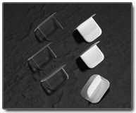
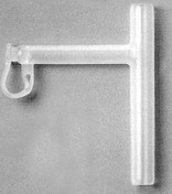
Keels come in different sizes. These are used to prevent adhesions, restenosis and web formation. This is particularly useful following glottic surgeries. It keeps both the anterior and posterior commissures separated thereby preventing adhesions. These keels are made of teflon, silastic or titanium. These materials are rather inert and do not irritate the soft tissues.
Keels should be long enough to extend from the crico thyroid membrane to 2 - 3 mm above the posterior commissure of the vocal folds. Superior portion of the stent should make a 120 degree angle to minimize granulation tissue formation at the level of posterior commissure / petiole of epiglottis. Keels can be placed endoscopically or through cricothyroidotomy. It is held in position by placing heavy sutures through the cricothyroid membrane. Ideally, these keels are left in place atleast for a period of 4 - 6 weeks. Removal of keel should ideally be performed under general anesthesia.
Stents:
Indications for the use of internal stenting are as follows:
1. Maintenance of the lumen after reconstruction in the absence of inadequate cartilagenous support.
2. Support for cartialge / bone grafts
3. To separate opposing raw mucosal surfaces during the healing porcess
- Note: If the stent is used to only allow mucosal healing / to allow graft take, it can be removed within 2 - 3 weeks. Prolonged use of stents i.e for more than a year is advisible only when there is grossly deficient laryngeal framework. Here in it is ideal for the scar tissue to develop around the stent.
Types of stents used:
Firm stents are used when splinting is required.
Solid stents are used if aspiration is a problem.
Soft hollow stents are used when use of voice is desirable.
Aboulker stent:
This is one of the oldest stents to be developed. It was first introduced in 1960. This stent is commonly used to stabilize tissues following laryngotracheal reconstruction in children with subglottic stenosis. This stent is shaped like a cigar and is made of teflon. Major advantage if this stent is that it causes very little irritation. The base of epiglottis can be irritated with formation of granulation tissue.
Montgomery stent:
This stent is made of silicone. It has a long central lumen and a smaller lumen projecting from the side of the stent at an angle of 90 degrees. This stent was first developed in 1965. This stent is used to stent the larynx and trachea after reconstruction for areas of malacia and stenosis. This stent is soft and can be retained for at least 1 year. Its main advantage is that it helps the patient to speak. It is more prone to crusting. It is contraindicated in children under the age of 2 as its lumen size is too narrow, and can be easily blocked.
Silastic sheet / Swiss roll:
This was first used by Evan's in 1977 for laryngotracheoplasty. The silastic sheet is rolled up and inserted into the larynx and upper trachea. It is stabilized in place by a stay suture. This roll has a constant tendency to unravel, thus producing steady pressure on the mucosa. This pressure has a beneficial effect by obliterating dead space allowing mucosa to regenerate.
Metal stents:
These stents are easy to use in distal trachea and bronchi. They are enmeshed and hence do not obstruct the bronchi. These stents include Palmaz and Streker stents. These stents are introduced via a bronchoscope.
Complications of stents:
1. Local infections
2. Mucosal ulceration
3. Granulation tissue formation
4. Stenosis


