Role of imaging in Otology
Contents
Introduction:
The past decade has ushered in a revolution in imaging techniques, the fall out of which is the ease with which we are able to see the inside of the body from varying planes and angles. It is a quantum leap when compared to the only radiological tool of yester years i.e. plain xray both mastoids Lateral oblique view (LAW'S view). The Law's view helped us only to compare the pneumatisation of the temporal bone on both sides, in addition to identifying the presence of cavity in the temporal bone. The advent of CT scan and MRI scan has helped us to pick up acoustic schwannomas at a very early stage.
Available imaging tools:
1. CT scan
2. MRI scan
3. Functional imaging
CT scan:
Synonyms: Computer assisted tomography, CAT scan
CT was invented in 1972 by British engineer Godfrey Hounsfield of EMI Laboratories, England, and independently by South African born physicist Allan Cormack of Tufts University, Massachusetts.
This imaging modality includes a sectional tomography, where in the body is radiographed in various sections. These sections are processed by a computer to generate three dimensional pictures. The data produced by a CT scanner can be manipulated through a process known as windowing. The process of windowing demonstrates the various structures based on their ability to block the X ray beams. The images obtained can be reformatted using a computer to provide saggital views of the structure being examined.
Contrast CT scanning:
Use of contrast agents help in assessing the nature of the tumor by assessing the vascularity of the mass. To study the vasculature of the mass intravenous contrast agents are injected just before the scanning process is to start. This produces temporary increase in the density of the arteries, the capillary-perfused parenchyma, and, finally, of peripheral veins. This is called contrast enhancement and is extremely useful for identifying vascular lesions. The intravenous contrast material is excreted renally, and therefore the renal collecting systems and ureters will be shown to be opacified on delayed CT scans.
Commonly used contrast preparations are iodine based, hence is contraindicated in patients with known allergy to iodine based drugs. Common drug used as contrast agent is Ultra vist, Angiograffin.
Advantages of CT scanning in otology:
1. It clearly demonstrates the intra cranial complications of suppurative otitis media.
2. It clearly delineates intracranial extension of tumors such as glomus jugulare / acoustic neuromas.
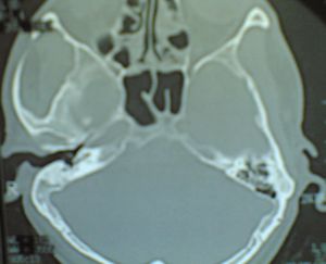
Routine CT views for petro mastiod area is axial with 1 - 2 mm cuts. These cuts are viewed with a wide window settings i.e. 3000 - 4000 H.U. (Bone window).
Caution:
Great care must be taken to limit radiation dose to the eye lens, cornea etc. This can be achieved if the scan is performed in a plane 30 degrees to the orbito meatal line. In this plane the globe is mostly below the part sectioned.
High resolution CT scan (HRCT):
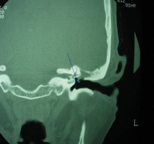
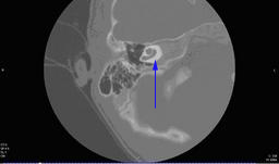
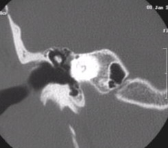
High-resolution computed tomography (HRCT) provides excellent contrast between osseous structures, air and soft tissue in conjunction with high spatial resolution. Therefore, thin-section HRCT with bone window setting is the method of choice for the examination of the middle ear structures. It is indicated in cases of acute / chronic inflammatory conditions of middle ear, cholesteatoma / tumors of middle ear. It clearly delineates bony walls and gives excellent resolution resulting in even minor erosions of ossicles, scutum to be clearly seen. It also depicts clearly the middle ear ossicles, and hence can be used to assess the position of ossicles postopertively after ossiculoplasty. HRCT can be used to identify congenital malformations involving cochlea. This will be helpful before performing cochlear implants in these patients.
It is also useful in identifying high jugular bulb.
A common condition labyrinthitis ossificans (ossification of the cochlea) can cause problems during cochlear implant procedures. These can be easily identified during a routine HTCT examination. Ossification of cochlea makes the procedure of cochlear implant a little difficult because the round window through which the implant is normally introduced is totally ossified, hence a third window will have to be drilled for optimal positioning of the electrode.
Magnetic resonance imaging:
The MRI scan uses magnetic and radio waves for imaging. The patient is not exposed to X rays. The patient is made to lie inside a large cylinder shaped magnet. Radio waves 10,000 to 30,000 times stronger than the earth's magnetic field are sent through the body. These radio waves affects the body's atoms forcing the nuclei into a different position. When these nuclei move back into their original positions they send out radio waves of their own which is picked up by the scanner, which is then converted by the computer into images.
Since our body mostly is made up of water, the nucleus of hydrogen atom which is a constituent of water is made use of in generating images of body. Using MRI scanner it is possible to image any part of the human body. The tissue which has very little hydrogen atoms (Bone) appear dark while tissues which have large number of hydrogen ions like fat appear much brighter. By changing the frequency of the radio waves it is possible to gain information about different types of tissues.
MRI scan will give clear images of tissues surrounded by bone i.e. brain and spinal cord. Traditional MR imaging comprises of non enhanced T1 and T2 weighted images, followed by gadolinum enhanced T1 weighted study.
T1 weighted images:
The term T1 indicates the time constant. It is the time taken by the body's atom to realign itself after being disturbed by radiowaves applied in a longitudinal axis. Non enhanced T1 weighted images in axial and coronal planes, identifies bone marrow, fat, and subacute hemorrhage as high (bright) signal areas. Gadolinum is used as a contrast agent to enhance images in MRI. Gadolinium-enhanced T1-weighted imaging in the same planes demonstrates enhancement when a neoplastic, vascular, or inflammatory process is present.
T2 weighted images: This is again another time constant. Here the radio waves are introduced in a transverse plane, and the time taken by a certain percentage of tissue nuclei to realign and get back into position is expressed as T2 weighted images. Standard T2-weighted studies identify intra-axial disease, such as brainstem tumor, stroke, and multiple sclerosis.
MRI signal characteristics:
Fat is brighter on T1- weighted imaging, infection or inflammation intensifies from T1- to T2- weighted imaging, and cerebrospinal fluid (CSF) turns from black to white from T1- to T2-weighted imaging. Bone appears dark on MR imaging.
Almost all the ossicular prosthesis used in the middle ear are non ferromagnetic except for Mcgee's stapes prosthesis. It is safe to perform MRI in patients with middle ear implants except Mcgee's prosthesis. Contraindications to MR imaging include pacemakers and aneurysm clips that are ferromagnetic.
MR imaging in patients with cochlear implants: The safety of patients who have undergone cochlear implant depends on the type of implant used. If imagining is done under low frequency i.e. 1 tesla imager, then it is safe. As a rule of thumb any MR imagining in a patient who has undergone a cochlear implant must be deferred unless and until it is absolutely essential, then too the manufacturer of the implant must be contacted regarding the safety of the procedure before imaging.
High resolution magnetic resonance imaging: is a recent innovation in imaging technique. This technique produces a heavily T2 weighted image. This helps in better visualisation of membranous labyrinth, and internal acoustic meatus. The fluid signal of the membranous labyrinth also appears bright, allowing for similar high-resolution examination of labyrinthine anatomy and abnormalities.
HRMR imaging has replaced HRCT as the preferred preoperative imaging study before cochlear implantation. It can verify the patency of cochlea, as well as to confirm the presence of cochlear nerve and its size. Without the presence of cochlear nerve surgery is not going to be successful. This study is especially beneficial if there is a history of meningitis and subsequent cochlear scarring is suspected.
3DFT-CISS: Three dimensional Fourier transformation constructive interference in steady state is one of the newest high resolution magnetic resonance imaging applications. Eventhough this study generates images (thin sections) in a single plane they can be reformatted with the help of computers into various planes. This imaging modality helps in identifying each and every nerve inside the internal acoustic meatus. Infact the origin of even the smallest acoustic neuroma can be identified.
This type of imaging is superior to the conventional MR imaging because:
1. The scanning time is fast
2. Gadolinum need not be injected for enhancement
3. Cheaper than conventinal MR imaging
4. Demonstrates clearly the membranous labyrinth, hence could be used to study patients with sensori neural hearing loss.
Disadvantages:
1. The screening field is limited
2. It is unable to demonstrate inflammatory lesions
Functional Imaging:
This type of imaging allows for measurement of cortical acitivity in response to a specific stimuli. The imaging modalities include:
1. Functional MRI (fMRI)
2. PET scan
3. SPECT scan
Functional MRI scanning: Is the recently developing scanning modality. MRI scanner is used to measure the hemodynamic activity related to the neural activity in brain and spinal cord. This procedure utilises the unique feature of hemoglobin where in it is diamagnetic when oxygenated and para magnetic when deoxygenated. These signals can be recorded as blood oxygen level dependent contrast (BOLD). Higher signal intensities can be recorded if the concentration of deoxyhemoglobin decreases. This leads to a higher BOLD value. The higher the tissue activity lower is the concentration of oxyhemoglobin, lesser is the value of BOLD.
Advantages of fMRI in otology are:
1. It can be successfully used in patient screening before cochlear implant surgery
2. It provides better spatial resolution
3. There is no need for contrast enhancement
4. This can be easily performed by stimulating the auditory center
PET scan: Positron emission tomography
This scanning modality produces a three dimensional map of the functional part of the body. To conduct the scan, a short-lived radioactive tracer isotope, which decays by emitting a positron, which also has been chemically incorporated into a metabolically active molecule, is injected into the living subject (usually into blood circulation). There is a waiting period while the metabolically active molecule becomes concentrated in tissues of interest; then the research subject or patient is placed in the imaging scanner. The molecule most commonly used for this purpose is flurodeoxyglucose (FDG), a sugar, for which the waiting period is typically an hour. This scan is hot when the tissue is highly active metabollically.
SPECT: Single photon emission computed tomography. This imaging is performed using gamma rays. It is performed using a gamma camera to acquire multiple 2 D images also known as projections. These images are fed in to a computer which reconstructs a 3 dimensional image. This type of functional imaging is helpful in evaluating patients after a cochlear implant. Since a gamma camera is used, there is no adverse effect due to the presence of an implant.
Role of imaging in studying cerebellopontine angle and internal acoustic meatus:
Imaging of this area is performed as a screening test to rule out acoustic neuroma in patients with unilateral sensori neural hearing loss. Gadolinum enhanced magnetic resonance imaging clearly demonstrates even the smallest of neuromas in this area. High resolution MRI can identify tumors of even 1mm size. If high resolution MRI produces a negative result it can be safely assumed that acoustic neuroma is not present. This scanning modality also clearly visualises the site of origin of the tumor. Since schwanomas are slow growing tumors, growth rate of vestibular schwanomas can be assessed by serial high definition MRI taken every 6 months after the initial diagnosis.
Post operative MRI scans can be taken 6 months after surgery to rule out residual disease.
HRCT can pick up acoustic neuromas of 2 cms diameter. It can be performed if MRI scanning is contraindicated in some patients.
Acoustic neuromas should be differentiated from meningiomas in this area. Meningiomas infact gives the same MRI imaging signal characteristics as schwanomas. In meningioma there is enhancement of adjacent dural trail, and the intracanalicular component of the mass is generally absent. Calcifications are commonly seen in meningiomas. Meningiomas commonly arise from cerebello pontine angle.
Epidermoid tumors present in the cerebello pontine angle provide a low (dark) signal on T1 weighted MRI, and a high (bright) signal on T2 weighted MRI. The signal intensity of the lesion is greater than that of CSF. Arachnoid cysts of this area show a more or less same signal intensity as epidermoid tumors, but they are difficult to differentiate from CSF.
MR angiography will help in the diagnosis of aberrent vessels in the CP angle area. They also clearly show dilated vessels as seen in aneurisms.
Study of facial nerve:
The choice of imaging modality for study of facial nerve depends on the suspected pathology and the segment of nerve involved. When the entire course of the nerve needs to be imaged then a conventional MRI will suite the bill. The complete examination of the nerve should be done from the brain stem up to the level of the parotid gland.
High resolution MR examination will help in the study of facial nerve within the internal acoustic meatus. But this type of scanning does not help in identification of Bell's palsy. The intra temporal portion of the facial nerve cannot be satisfactorily examined by MRI, only a high resolution CT scan will give a clear picture of the intra tympanic course of facial nerve. Temporal bone fractures can be clearly shown only in a high resolution CT scan. In cases of suspected tumors involving the facial nerve HRCT and gadolinum enhanced contrast MRI will prove complimentary to each other. MR imaging is not indicated routinely for Bell's palsy. In the case of atypical Bell's palsy, then gadolinum enhanced MRI should be performed to rule out tumors involving the nerve. Normal MRI in patients with pure Bell's palsy will show enhancement without focal enlargement of the nerve.
CSF leaks involving the temporal bone: can be studied using HRCT. Contrast can be injected intrathecally before scanning to clearly show the site of leak.
Imaging Petrous apex area: This area is a very silent area. It provides very few early symptoms, hence imaging is the only way to pick up early lesions. Infact lesions of the petrous apex have been incidentally identified on routine scanning of the area. HRCT and gadolinum enhanced MR scanning help in identifying lesions of this area. HRCT clearly shows bony destruction if present in this area.
Cholesterol granuloma and epidermoid of the petrous apex can often be differentiated by using T1 weighted Magnetic resonance imaging. Cholesterol granuloma appears bright on T1 weighted signal where as the epidermoid cyst appears dark on T1 weighted imaging.
Imaging jugular foramen area: To examine jugular foramen area both HRCT and gadolinum enhanced MRI scan should be performed. In glomus jugulare the bone erosion seen in HRCT resembles a moth eaten appearance.
Examination of labyrinth: Congenital anamolies involving the labyrith can be studied using HRCT in combination with HRMRI. A perilymph gusher may be predicted before stapedectomy by performing high resolution MRI. This is so when the CSF signal is identical to the perilymphatic fluid signal at the internal acoustic meatus.
Patients with complicated middle ear inflammatory disease will have to under go MRI scanning to rule out intra cranial complications. This scanning modality also helps in the identification of sigmoid sinus thrombosis.
External auditory canal: Imaging of external canal is performed only in cases of refractory otitis externa. Scanning will help in identifying minor bone erosions which could occur in patients with malignant otitis externa.


