Recent nomenclature changes in classification of CSOM
Contents
Introduction:
Chronic suppurative otitis media, its classification, and definition has undergone changes. Chronic suppurative otitis media has been rechristened to chronic otitis media. The term chronic otitis media implies a permanent abnormality of the pars tensa / pars flaccida as a result of acute otitis media, negative middle ear pressure or otitis media with effusion.
Chronic otitis media simply does not mean accumulation of pus in the middle ear cavity. It can be subdivided into active chronic otitis media (active com) and inactive chronic otitis media (inactive com).
Active COM:
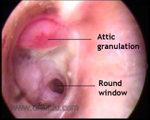
In this condition there is inflammation of middle ear mucosa associated with accumulation of pus. There may also be associated mastoiditis. Active COM can be subdivided into active mucosal COM and active squamosal COM.
Active mucosal COM:
Ear drum in these patients will be perforated. The middle ear mucosa may undergo polypoidal changes causing "aural polypi". It is also important to realize that inflammatory changes in this disorder is not confined to the middle ear alone, the whole of the middle ear cleft is involved. Simple closure of the perforation without removal of infected middle ear mucosa and granulations from the mastoid cavity is fraught with failure to control the disease.
Active mucosal COM is often associated with resorption of parts or whole of ossicular chain. This could be due to resorptive osteitis. The ossicles affected typically show hyperemia with proliferation of capillaries and prominent histiocytes. Long process of incus gets eroded commonly, followed by stapes crurae, body of incus and manubrium in that order.
Active squamous COM:
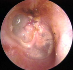
This condition is otherwise known as unsafe ear or cholesteatoma. This condition is commonly associated with retraction of pars flaccida / tensa that has retained squamous epithelial debris. There is also associated inflammation of middle ear mucosa, production of pus, and erosion of ossicles. This condition is commonly associated with intracranial complications.
Inactive Chronic otitis media:
In this condition the middle ear mucosa is relatively healthy. The mastoid cavity also appear healthy. These patients may slip into active phase rather easily because of the existing pathology. Inactive chronic otitis media can further be subdivided into Inactive mucosal chronic otitis media and Active squamous chronic otitis media.
Inactive mucosal chronic otitis media:
This condition is always associated with dry perforation of the ear drum. There is permanent perforation of the pars tensa, but the middle ear and mastoid mucosa are not inflamed. The drum remnant around the perforation is always healthy. The rim of the perforation is thickened due to proliferation of fibrous tissue. Squamous epithelial cells from the external auditory canal does not migrate into the middle ear cavity in this stage because the annulus of the ear drum is intact and it prevents this migration. These patients benefit from myringoplasty.
Inactive squamous epithelial chronic otitis media:
These include retraction pockets, atelectasis and epidermization. Negative middle ear pressure can cause retraction of tympanic membrane. A retraction pocket consists of an invagination into the middle ear space of part of the ear drum. These retraction pockets may be fixed when it is adherent to structures in the middle ear or free when it can move freely medially or laterally depending on the state of inflation of the middle ear. "Epidermization" is a more advanced type of retraction and it refers to replacement of middle ear mucosa by keratinizing squamous epithelium without retention of keratin debris. The area of epidermization may involve part or all of the middle ear cavity. Epidermization often remain quiescent and does not progress to cholesteatoma or active suppuration. So epidermization per se is not an indication for surgical intervention.
Healed chronic otitis media:
In this stage the perforated ear drum has managed to heal itself. Loss of lamina propria or the tympanic membrane due to atrophy or failure of complete healing leads to a 'dimeric' membrane that consists of epidermis and mucosa only. Such thin membrane is more prone to retraction if there is negative middle ear pressure.
Tympanoslcerosis is also another form of healed ear drum. It refers to hyaline deposits of acellular material visible as whitish plaques in the tympanic membrane or as white nodular deposits in the submucosal layers of the middle ear on otoscopy. Tympanosclerosis is the end result of a healing process in which collagen in fibrous tissue hyalinizes, loses its structure and become fused into a homogenous mass. Calcification and ossification may occur to a variable extent.


