Pseudocyst of Pinna
Contents
Introduction:
This condition involves the Pinna and can frequently recur even after successful treatment. It goes by various names i.e. intercartilagenous cyst, endochondral Pseudocyst and idiopathic cystic chondromalacia. This condition was first described by Engel in 1966.
Clinical features:
1. Presents as painless, spontaneous dome shaped cystic swelling on the anterior surface of auricle.
2. This condition is predominantly seen in adult males
3. It is uncommon before 20 and after 60 years of age.
4. Majority of these cysts are found in the scaphoid and triangular fossae of the pinna
5. Majority of these cysts have been reported in Chinese. Chinese have attributed this problem due to the firm pillow they use to sleep. Studies have not demonstrated any racial differences.
6. Right ear is more commonly affected than the left. This has been attributed to the habit of majority of individuals to sleep on their right side.
Histology:
Histologically, pseudo cyst of pinna is characterized by an intercartilagenous cavity which lacks an epithelial lining hence the term pseudocyst is used to denote this condition. During later stages the cartilage may show evidence of intercartilagenous fibrosis and granulation tissue formation. Perivascular infiltrates of lymphocytes indicate that this condition is predominantly inflammatory in nature.
Needle aspirate from the lesion will demonstrate the presence of straw colored fluid whose concentration of protein is more or less similar to that of plasma.
Etiopathogenesis:
Etiology of this conditions is largely unknown. Many theories have been proposed to explain this condition. They include:
1. Engel's theory: Engle hypothesized that release of lysosomal enzymes from local chondrocytes caused progressive dilatation and formation of intracartilagenous cavity. Studies have demonstrated that there is no increase in lysosomal activity in the aspirated fluid. Electron microscopic examination of chondrocytes have also failed to demonstrate increased numbers of lysosomes in them.
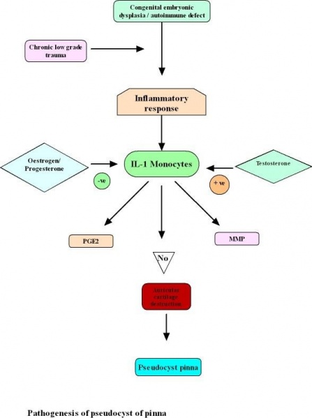
2. Congenital embryonic dysplasia theory: This theory proposes that dysplasia of the auricular cartilage could be a factor in the development of pseudocyst of pinna. The auricle develops from the fusion of 6 tubercles surrounding the first and second branchial arches. During this complex folding and fusion processes residual tissue planes may be formed within the mesenchyme that gives rise to the pinna. These tissue planes can be reopened later leading on to the formation of pseudocyst. The presence of this potential intercartilagenous space can be identified histologically in patients.
3. Chronic low grade trauma: This has been attributed to be one of the causes for formation of pseudocysts of pinna. Predominant involvement of right ear is accounted by the fact that most of the patients tend to sleep on their right side cause chronic low grade trauma to their pinna. Wearing of motor cycle helmets, headphones to listen to music can also cause chronic low grade trauma to pinna. According to Choi repeated minor trauma led to over production of Glycosaminoglycans. These gyclosaminoglycans form microcysts within the cartilage. These cysts later coalesce to form pseudocyst of the pinna. Evidence that point towards this theory of chronic mild trauma is the presence of hemosiderin and LDH within the pseudocyst fluid. LDH – 4 and LDH – 5 are the major LDH isoenzyme components of auricular cartilage. These two components have been found to be elevated in the pseudocystic fluid.
4. Inflammatory theory of LIM: LIM identified the presence of perivascular infiltrates of lymphocyts in the connective tissue just superficial to the anterior segment of the auricular cartilage examined. On this basis he postulated that inflammation could play a vital role in the pathogenesis of this condition. He also believed that these inflammatory cells release cytokines which caused cleavage of the auricular cartilage.
5. Autoimmune theory – Autoimmunity can stimulate inflammatory response leading to the formation of pseudocyst. Antinuclear antibodies have been detected in the cystic fluid lending credence to autoimmune theory.
Interleukin I is the cytokine responsible for inflammatory reaction. Moncytes and macrophages are the primary source of Interleukin I. It stimulates the production of immune mediators like Prostaglandin E2, nitric oxide, cytokines, chemokines and adhesion molecules that are involved in cartilaginous inflammation. In addition to these substances IL I also stimulates synthesis of matrix metalloproteinases (MMP) and other enzymes involved in cartilage destruction of cartilage.
Studies have shown that oestrogen and progesterone inhibited the production of IL I by peripheral monocytes, testosterone on the other hand increased the production of IL I by monocytes. This accounts for the reason that pseudocyst of pinna is common in males.
Differential diagnosis:
Pseudocyst of pinna can be confused with:
1. Relapsing polychondritis 2. Chondrodermatitis nodularis helicis 3. Cauliflower ear 4. Subperichondrial hematoma
The differentiating point between Pseudocyst pinna and the conditions mentioned is that pseudocyst is in the intercartilagenous plane while all these conditions occur in the subperichondrial plane.
Management:
1. Antibiotics 2. Anti inflammatory drugs 3. Steroids (systemic / intralesional) ??
Wide bore needle aspiration – The fluid from pseudocyst can be aseptically aspirated using a wide bore needle. Compression dressing is applied in order to avoid recurrence due to fluid reaccumulation. POP can also be used as compression dressing.
Button surgery: Incision and drainage always was associated with a high recurrence rate. This procedure is performed under local infiltrative anesthesia. A helical incision is made, and the skin flap is elevated well beyond the anterior cartilage segment of the pseudocyst. The anterior wall of the cyst is excised along the margin. After the straw colored fluid is drained completely the posterior wall is curetted. After obtaining hemostasis, similar sized buttons (sterile) is sutured on the anterior and posterior surfaces of the pinna providing compression at the operated area. This helps in preventing recurrence in these patients. No other dressing is essential. The patient is put on antibiotics and anti inflammatory drugs for a week after which the suture is removed along with the shirt button.
