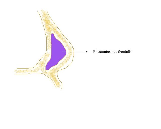
Pneumosinus dilatans of para nasal sinuses
Introduction:
Pneumatization of paranasal sinuses are highly variable. These sinuses are lined by normal mucosa. Rarely pneumatization extends beyond the confines of the frontal bone, causing swelling of the sinus (Pneumosinus dilatans frontalis). The lining epithelium is normal in these patients. About 20% of pneumosinus dilatans cases are associated with arachnoid cysts, meningioma, fibrous dysplasia, acromegaly.
MRI scans are thus indicated when CT scan shows suspicious lesions.
Clinical features:
1. Deformity
2. Frontal pain
3. Barotraumas
4. If sphenoid sinus is affected visual disturbances are common
Aetiopathogenesis:
Aetiology is still unclear. It could be caused by spontaneously discharging frontal sinus mucocele. A valvular mechanism (ball valve type) has been postulated to be a cause for frontal sinus mucocele due to large anterior ethmoidal cell, and mucosal swelling of the frontal recess area. Increased pressures have been documented in these affected sinuses. Sometimes paranasal sinus pneumatization is stimulated by growth and sexual hormones. These hormones regulate the activity of osteoblasts and osteoclasts.
Treatment:
Surgery is the ideal treatment of choice. In frontal sinus pneumatosinus, horizontal bone strips are removed after elevating the anterior wall of the frontal sinus. If obliteration is required, the sinus mucosa is diligently removed using a microscope.

