Pneumatic otoscopy
Contents
Introduction:
This examination procedure determines the ear drum mobility in a patient in response to pressure changes in the external auditory canal. Normal ear drum moves inside when the pressure in the external auditory canal is increased, and outside if the pressure is reduced. This to and fro movement of the ear drum in response to pressure changes in the external auditory canal can be visualized using a siegel's penumatic speculum or pneumatic otoscope.Reasons of immobile ear drum despite pressure changes in the external canal:
1. Fluid in the middle ear cavity – reduction / restriction of ear drum mobility in response to pressure changes in the external ear
2. Perforated ear drum
3. Adhesive otitis mediaPneumatic otoscopy has a high predictive value for otitis media with effusion even in the presence of morphologically normal ear drum.
History:
History of otoscopy makes an interesting reading. Early otoscope was nothing but a tong shaped specula. This device was first described in French literature by Guy de Chauliac in 1363. This same speculum was also used to examine the nasal cavity. In 1838 Ignaz Gruber redesigned the tong shaped aural speculum into a funnel shaped one. The currently available Gruber's aural speculum was designed by him. In 1864 E. Sigel of Germany designed the Siegel's pneumatic aural speculum which is still being used today to study mobility of ear drum. He should be considered as the father of penumatic otoscopy. The model proposed by Siegel enabled otologist to apply pneumatic pressure over the ear drum. It was Politzer who popularized pneumatic otoscopy as a routine examination procedure in 1909 .
Indications:
In paediatric age group pneumatic otoscopy helps to identify otitis media with effusion. This is infact a quick and painless way of identifying a child with otitis media with effusion. Otitis media with effusion is a common childhood condition causing conductive deafness and delayed speech.
1. Pneumatic otoscopy also helps in identifying retracted ear drum
2. Also helps in differentiating retracted ear drum from a large central perforation
3. Can be used to test eustachean tube function
4. Helps in the assessment of landmarks of ear drum
5. Used to differentiate otitis media with effusion from acute otitis mediaWhen the tympanic membrane is retracted due to negative middle ear pressure, the ear drum movement is greater when the bulb is released than when the bulb is compressed.
Equipments necessary for pneumatic otoscopy:
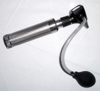
1. Otoscope with pneumatic attachment
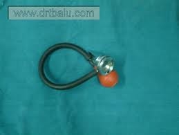
2. Siegels speculum
3. Either one of the above two said equipment can be utilized for this purpose.
Procedure:
A snuggly fitting aural speculum is chosen for performing this test. This speculum can be attached to an penumatic otoscope / siegel's pneumatic speculum holder. The speculum is gently inserted into the external auditory canal of the patient. This speculum straightens the external canal bringing the ear drum into full view of the examiner. The optical lens system present in the otoscope / siegel's speculum provides an enlarged image of the ear drum. The bulb of the pneumatic apparatus is pressed and released and the movement of the ear drum in relation to the pressure changes brought about at the external auditory canal is observed.

