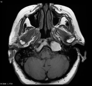
Petrous apex cholesterol granuloma
Cholesterol granulomas represents the most common cystic lesion of petrous apex. Cholesterol granulomas of middle ear cavity was first described by Manasse in 1917. Shambaugh in 1929 described idiopathic hemotympanum as a cause to the development of cholesterol granuloma of middle ear. This is actually a special type of granulation tissue affecting the middle ear cavity and is prone to bleeding. It is hence a frequent cause of hemotympanum.
These lesions typically affect young to middle aged individuals. These patients often present with a h/o chronic otitis media. This disorder does not manifest any gender prediliction. Temporal bone cholesterol granuloma are known to appear in three forms:
Associated with chronic otitis media
Idiopathic hemotympanum
Localised disease of middle ear cavity, mastoid antrum, external auditory canal and petrous apex.
Clinical presentation:
It depends on the location of the lesion. If it is present in the middle ear cavity then:
1. Conductive hearing loss 2. Dizziness 3. Cranial nerve dysfunction (7th nerve)
If it involves the petrous apex then it could present with:
1. Hearing loss (65%) conductive in nature
2. Vestibular symptoms (giddiness) 55% of cases
3. Head ache reported in nearly 30% patients
4. Facial twitching in 24% patients
5. Facial paresthesia in 20% of patients
6. Otorrhoea 12% of patients
7. Diplopia 6% of patients
8. Facial weakness in 3% patients
Petrous apex cholesterol granuloma can be divided into:
Type I:
In this type due to vestibulocochlear involvement sensorineural hearing loss and tinnitus are the commonest symptom followed by vertigo and dizziness.
Type II:
In this type the symptom is caused due to spread to the upper part of the petrous apex causing middle and posterior cranial fossa dural irritation. Head ache and facial pain are the commonest symptoms.
Type III:
Symptoms in this type are caused by compression of trigeminal / abducens nerve indicating compression of Meckle's cave region. Rarely recurrent otitis media could also occur as a result of eustachean tube compression.
Cholesterol granuloma involving the petrous apex are considered to be more aggressive than their counterparts involving mastoid cavity and middle ear.
If it involves the mastoid then
1. It could be asymptomatic
2. Headache
Pathology:
It can involve any portion of the aerated temporal bone. The masoid air cell system happens to be the most common location.
Pathogenesis is actually controversial. There are currently two theories.
Obstruction vacuum theory :
Eustachean tube dysfunction is considered to be the root cause for this problem. It causes mucosal oedema with repeated episodes of bleeding.
Exposed marrow theory:
In this theory hyperplastic mucosa invades the underlying bone and exposes the bone marrow. This in turn causes bleeding.
Histology:
It is composed of yellowish brown fluid. This fluid contains:
Cholesterol crystals - This accounts of the high T1 and T2 signal
Multinucleated giant cells
Red cells and their break down products
Hemosiderin
This is surrounded by fibrous connective tissue with fragile blood vessels that could easily rupture.
Radiology:
CT imaging:
Shows expansile well marginated lesion with thinned overlying bone. Bone may be dehiscent when the lesion is large. Faint peripheral enhancement when contrast is used could be seen. Appearance of the lesion is determined by its location. When a cholesterol granuloma is located in the petrous apex then it could appear to be more aggressive with evidence of bony erosion and extension into the carotid canal or cerebello pontine angle. When it is located in the middle ear the associated erosion is not clear.
MRI:
T1 weighted image:
Shows high signal intensity due to cholesterol component and methemoglobin. The rim of the lesion could have a low signal because of the presence of hemosiderin in the rim area and thinning of the adjacent bone.
T2 weighted image:
Shows central high signal with peripheral low signal due to hemosiderin rim
Thinning of adjacent bone
Does not attenuate on FLAIR
Fat suppression: Remain high signal
T1 contrast using Gadolinum:
There is no central enhancement and peripheral enhancement may be difficult to identify high T1 signal which is not saturated (not adipose tissue).
Diffusion weighted coefficient and apparent diffusion coefficient:
Shows no restricted diffusion
Diffential diagnosis:
1. Normal asymmetry of fatty marrow / pneumatization
2. Middle ear effusion
3. Effusion of pneumatized petrous apex
4. Cholesteatomas
5. Base of skull tumors like chondrosarcoma / metastatic lesions
6. Mucoceles
7. Thrombosed internal carotid artery aneurysm
Treatament:
If symptomatic surgical excision is requried. Excision should include the complete cyst wall. The surgical approach chosen depends on the site of the lesion and degree of hearing loss. Mastoidectomy is commonly resorted to procedure. Recurrence rates are very high.
Aggressive cholesterol granuloma requires surgery that would drain / resect the disease, preserve hearing if possible and preserve cranial nerve and carotid artery. CSF leak should also be prevented.
Fenestration procedures:
Initially this procedure was used to drain the cyst. Drainage was performed via tubes inserted for this purpose. This procedure was not successful because it did not drain the cyst completely.
Middle cranial fossa approach:
This approach is recommended for managing aggressive petrous apex cholesterol granuloma that has spread towards the middle cranial fossa. This procedure is used for complete removal rather than simply drain the cyst. This approach is preferred when poor hypotympanic pneumatization and the location of the cyst makes infracochlear approach rather difficult. This approach provides good access to remove all the cyst except those cysts that have spread inferiorly or those that encircles the carotid artery.
Infralabyrinthine & infracochlear apprach:
This approach is commonlly used in patients with normal hearing. Patients with highly placed jugular bulb are not ideal candidates for infralabyrinthine approach. Infracochlear hypotympanic approach is the most common approach used to access petrous apex. This is a more conservative procedure to drain, decompress and ventilate the cyst. This procedure is safer and simpler than the middle cranial fossa approach because it avoids injury to the Greater superfical petrosal nerve, facial nerve and ET and preserves the carotid artery and inner ear. Pre op CT scan should be obtained to show good penumatization between the cyst wall and hypotympanum.
Transcochlear approach:
This is the best approach to the petrous apex in deaf patients. It provides good exposure and control of the carotid artery for large lesion.
Trans spenoidal endoscopic surgical approach:
This procedure can be used for cysts reaching the sphenoid sinus. There should be space between the cyst wall and the lateral wall of paralegal internal carotid artery. It is contraindicated in patients with cysts hidden behind the internal carotid artery. Major disadvantage of this procedure is the possibility of recurrent stenosis occurring at its opening.
In patients with middle ear, mastoid, or External auditory canal lesions then tympanomastoidectomy with appropriate reconstruction procedure should be used.
Asymptomatic small lesions are managed conservatively with regular clinical and radiological follow up.
In open surgical procedures success can be achieved in about 90% of patients with symptomatic improvement. Recurrence rate is about 10% and complication rates in the range of 25% (which could be very high). Common complication of them happens to be the hearing loss produced due to surgery.
Post op CT image will reveal air within petrous apex indicating good areation of the cyst which has been surgically drained. If the cyst is refilled with serous fluid or scar tissue then CT imaging may not be sufficient to differential these lesions. MRI is indicated in these patients. Successful management would show up in MRI as decrease in signal intensity in T1 weighted images with reduction in the size of the lesion.
MRI image showing cholesterol granuloma involving petrous apex
