Mucocele
Contents
Definition:
These are epithelial lined, mucus containing sac which completely fills the sinus cavity. They are also capable of expansion. This condition is different from that of a blocked sinus cavity which simply contains mucus.
Sites of occurrence:
The fronto ethmoidal region is the most common site, followed by sphenoid sinus. Mucoceles involving the maxillary sinus are pretty rare. The fronto ethmoidal region is commonly involved because of its complex drainage pattern when compared to that of maxillary and sphenoid sinuses.
Etiology:
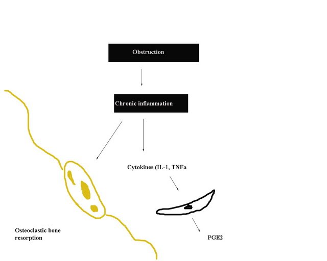
Mucoceles are fairly uncommon, and in 1/3 of cases occur without any predisposing factors. It is so slow growing that there may be a considerable time lag between the initiating factor and the clinical presentation of mucocele. In the case of surgery or trauma the average time lag could exceed two decades. Acute infections have a somewhat lesser time lag (2 years). Mucoceles are thought to arise as a consequence of obstruction and inflammation.
Pathogenesis:
Three main theories of pathogenesis are available.
They are: 1. Pressure erosion 2. Cystic degeneration of glandular tissue 3. Active bone resorption and regeneration Mucoceles are lined by pseudostratified columnar epithelium with squamous metaplasia. There may also be associated goblet cell hyperplasia. The cellular infiltrate present within the lining mucosa include components of both acute and chronic inflammation.
Clinical features:
Patients with frontoethmoidal mucoceles are first seen by the ophthalmologist because of proptosis. CT scan paranasal sinuses is diagnostic. Xray paranasal sinuses water's view will show exapansile mass inside the frontal sinus with loss of frontal sinus hausterations. This is one of the important diagnostic features of frontoethmoidal mucocele.Diagnostic nasal endoscopy may reveal an expansile mass presenting inside the nasal cavity. Mucoceles of maxillary sinus may expand into the nasal cavity producing nasal obstruction, or may erode the anterior wall of maxilla at the level of canine fossa causing a cheek swelling.Sphenoidal mucoceles due to its intimate relationship with orbital apex and cavernous sinus may present with signs of visual disturbances.Frontoethmoidal mucoceles will present as swelling of the orbit, causing proptosis. It may also erode the outer table of the frontal sinus causing a swelling over the forehead region.
Management:
Endoscopic sinus surgery is almost curative in all these cases.
Tip:
Whatever be the type of approach used, there is no necessity to reconstruct the areas where bone resorption has occurred. As long as the mucosal lining is intact, restitution of contour occurs rapidly. In young patients reossification can also occur.
