Keratosis obturans
Contents
Keratosis obturans:
is accumulation of desquamated keratin in the external auditory meatus. This should be differentiated from primary auditory canal cholesteatoma which is characterized by invasion of squamous tissue from the external ear canal into a localized area of bone erosion.
Pathology:
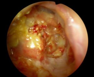
The keratin plug seen in keratosis obturans appears like a geometrically patterned keratin plug within the lumen of expanded ear canal. These keratin squames are shed from the complete circumference of the deep ear canal forming a lamina. It appears like onion skin.
Etiology:
Keratosis obturans is postulated to occur due to abnormal epithelial migration of ear canal skin. The movement of the surface epithelium appears to be reversed in these patients. (The surface epithelium over pars flaccida migrates downwards to the pars tensa and then moves inferiorly across the drum).
Keratosis tympanicum:
Is also caused by abnormal migration of squamous epithelium lining the deep portion of the external auditory canal. This condition is also associated with unilateral tinnitus.
Types of keratosis obturans:
a. Inflammatory type: This is caused due to acute inflammation involving the external ear canal. Viral infections commonly cause this problem. The inflammatory reaction involving the ear canal temporarily alters epithelial migration. This condition can only be cured by removal b. Silent type: In this type there is no predisposing acute infections involved. This condition is postulated to be caused by abnormal separation keratin that persists even after the removal, and will need repeated removals. c. Primary auditory canal cholesteatoma: Etiology is uncertain. It is commonly thought to be caused by trauma to the bone covering the external canal. This could also be caused by surgical trauma as in patients who have undergone stapedectomy. The piece of exposed bone in the external canal becomes infected and sequests. The lining epithelium migrates into this area causing the formation of cholesteatoma. This condition is characterized by ear pain which is dull and aching in nature. It is not associated with hearing impairment.
Keratosis obturans commonly occur in young patients.
Clinical features:
1. Severe ear pain
2. Mild / moderate conductive hearing loss
3. Associated bronchitis / sinusitis - common
On examination:
The ear canal appears to be widened, making the ear drum stand out. CT scan of temporal bones may reveal canal erosion and widening.
After surgical removal under general anesthesia the specimen must be sent for pathological evaluation to rule out malignancy.
Management:
1. Surgical removal under G.A.
2. Canal plasty is helpful in recurrent cases
3. Mastoidectomy should be performed in cases with primary cholesteatoma of external canal.
