Hemangioma parotid gland in an adult an interesting case report
History:
1. Swelling over right angle of jaw - 3 years - Gradual increase in size
2. No history of pain
On examination:
Soft globular swelling measuring 4 x 3 cms seen occupying the right angle of mandible. It was warm and compressible.
The skin over the swelling was free.
The swelling could be moved over underlying tissues.
Transillumination negative
Imaging:
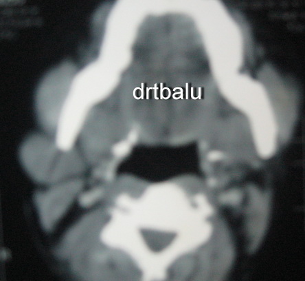
Axial CT of the area showed radiolucent hypodense globular mass attached to the lower pole of the parotid gland. Contrast CT showed contrast enhancement of the mass.
FNAC:
The tap turned out to be bloody in nature.
Management:
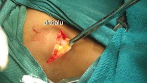
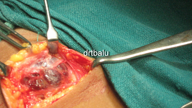
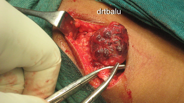
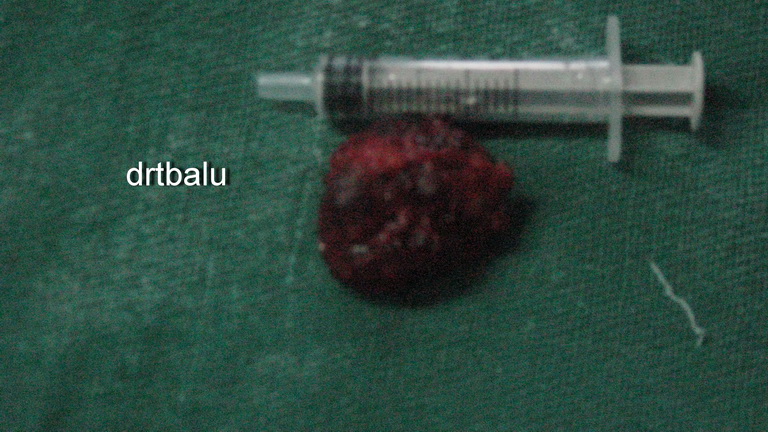
The patient was taken up for surgery under general anesthesia. The mass was removed in toto.
Histopathology report:
Consistent with cavernous hemangioma of parotid gland
Discussion:
Hemangioma of parotid gland is a rather common lesion in infants and children. It is rather rare in adults. Histologically this mass is composed of solid masses of cells with capillary vascular spaces. Normal salivary gland tissues are usually replaced by the mass. These masses are usually soft, warm and compressible and donot transilluminate. A period of wait and watch is usually followed. Indications for surgical intervention include rapid increase in size, and pain.
There is still a raging controversy whether hemagioma could be a true tumor involving blood vessels or a vascular malformation (Hamartoma).
These masses usually become prominent when the head is bent forward or when the patient lies horizontally (This is classically known as Turkey wattle sign). Rarely multiple phleboliths can be seen within the mass.
Peel & Gnepp classified hemangioma of salivary glands as follows:
1. Benign hemangioendothelioma
2. Cavernous hemangioma
3. True capillary hemangioma (rare)
There is absolutely no role for scerotherapy in these patients.

