Grommet insertion current trends
Introduction:
Myringotomy with grommet insertion was introduced by Poltizer of Vienna in 1868. He used this procedure to manage “Otitis media catarrhalis”. Soon it became the common surgical procedure performed in children.Indications:Bluestone and Klein (2004) came out with revised indications for grommet insertion which took into consideration the prevailing antibiotic spectrum.chronic otis media with effusion not responding to antibiotic medication and has persisted for more than 3 months when bilateral or 6 months when unilateral.
Recurrent acute otitis media especially when antibiotic prophylaxis fails. The minimum episode frequency should be 3/4 during previous 6 months / 4 or more attacks during previous year.Recurrent episodes of otitis media with effusion in which duration of each episode does not meet the criteria given for chronic otitis media but the cumulative duration is considered to be excessive (6 episodes in the previous year)
Suppurative complication is present / suspected. It can be identified if myringotomy is performed.Eustachean tube dysfunction even if the patient doesn't have middle ear effusion. Symptoms are usually fluctuating (dysequilibrium, tinnitus, vertigo, autophony and severe retraction pocket).Otitis barotrauma inorder to prevent recurrent episodes.
Problems with Grommet insertion:This procedure is not without its attendant problems. Common problems include:
1. Segmental atrophy of tympanic membrane
2. Tympanosclerosis
3. Persistent perforation syndrome (rare)
Before treating patients with otitis media with effusion the following factors should be borne in mind.Pneumatic otoscopy should be used to differentiate otitis media with effusion from acute otitis media.
Duration of symptoms should be carefully documented.
Children with risk for learning / speech problems should be carefully identified. Hearing should be evaluated in all children who have persistent effusion for more than 3 months.
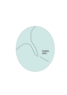
Grommet insertion can be performed under local anesthesia. Incision is made in the antero inferior quadrant of ear drum. The incision is given along the direction of radial fibers of the middle layer of ear drum. This causes minimal damage to the radial fibers. It also enables these fibers to hug the grommet in position.
The site of incision is shown. The drum is incised using a sickle knife. The opening is for drainage purpose and hence the incision is given against the course of middle fibrous layer of ear drum. If grommet is introduced for ventilation purpose then the incision should be given along a parallel line of the middle fibrous layer.


