Endoscopic management of frontoethmodial mucocele with intracranial extension
68 year old female patient reported to ENT out patient department with
1. C/O swelling left side of forehead - 2 years duration
2. Headache on and off - 2 years
3. Swelling over left eye - 1 1/2 years
The preoperative picture shows the patient having swelling over her left forehead with the left eye pushed downwards and outwards.
O/E
Vision normal in both eyes
CT scan of the patient clearly showed the swelling over left frontal sinus with erosion of the outer and inner tables. The mass is also seen to extend into the frontal lobe of the brain.
The clinical diagnosis was frontal mucocele with intracranial extension.
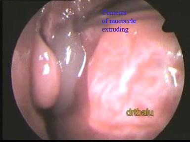
This patient underwent endoscopic decompression of the mucocele through the nasal cavity.
The major advantages of endoscopic approach are
1. The procedure has minimal risk
2. There is no scar
3. Intranasal drainage path can be created
4. Minimal complications
Surgical procedure:
Using a 4mm 0° nasal endoscope the surgery was performed. The complete surgery was performed under general anesthesia. On deroofing the agger nasi cell the contents of the mucocele started to extrude. The frontal sinus ostium was widened. When the scope was introduced through the widened frontal ostium the posterior table of the frontal sinus was found to be eroded. The frontal lobe of the brain was clearly visible. The brain can be identified by its characteristic pulsations coinciding with the patient's respiration.
Discussion:
A mucocele is an epithelium lined mucous containing sac. It usually develops when the sinus ostium gets obstructed by chronic sinusitis, polyps or tumors. These mucoceles are known to erode the bone and may involve the brain and orbit. It may also present as a forehead mass with proptosis as in this patient.
Classification of Frontal mucocele:
Frontal mucoceles have been classified into 5 types depending on its extent.
Type I: In this type the mucocele is limited to the frontal sinus only with or without orbital extension.
Type II: Here the mucocele is found involving the frontal and ethmoidal sinuses with or without orbital extension.
Type IIIa: In this type the mucocele erodes the posterior wall of the frontal sinus with minimal or no intracranial involvement.
Type IIIb: In this type the mucocele erodes the posterior wall with major intra cranial extension.
Type IV: In this type the mucocele erodes the anterior wall of the frontal sinus.
Type Va: In this type there is erosion of both anterior and posterior walls of frontal sinus without or minimal intracranial extension.
Type Vb: In this type there is erosion of both anterior and posterior walls of frontal sinus with a major intracranial extension.
Among mucoceles affecting the various paranasal sinuses frontal mucoceles are the most common (65%). Before the advent of CT scan x-ray paranasal sinuses was the only diagnostic tool available. X-ray would usually reveal the loss of normal haustrations found in the frontal sinus. Infact it was even considered pathognomonic.


