Clinical examination in laryngology
Examination of Ear Nose and Throat needs specialised illumination and instruments. Since these are cavities proper illumination, focussing of illumination special instruments are necessary.
Two types of illumination is used in otolaryngologcial examination:
1. Semi mobile illumination like the Bull's lamp
2. Mobile illumination like the Clair's head light, or cold light based head bands.
Bull's lamp: is a semi mobile source of illumination. It has a 100 watts milk white bulb which provide the source of illumination. This light is focussed with theplano convex lens placed in front of the bulb. Ideally the Bulls lamp is placed 6 inches above and behind the left shoulder of the patient, at the level of left ear of the patient.
The examiner and the patient must be conveniently be seated on adjustable stools. While using a Bull's lamp the examiner must focus the light using a head mirror to illuminate the patient.
The head mirror is a concave mirror. It has a hole in the centre. The approximate focal length of the mirror is about 10 inches. It has a plastic head band with a lever with 2 ball and socket joints. The joints are at right angles to each other.
Focusing the light with head mirror: The art of focusing the light to the desired spot comes only with practice. But there are certain basic steps which must be followed.
1. The patient sitting on the stool must be at the same level as the doctor.
2. The patient's legs must be placed to one side of the examiner.
3. The distance between the doctor and the patient must not be more than 8 inches (i.e. the focal length of the head mirror).
4. The mirror is fixed over the right eye in such a way part of the mirror touches the nose.
5. The mirror is adjusted in such a way that the right eye sees through the hole in the mirror. The mirror is adjusted while keeping the left eye closed and the right eye is kept open. Then both eyes are opened.
Clair's head light provides mobile illumination. The source of light is from a small 9 volt bulb. It is placed in front of a adjustable concave mirror. The mirror and the bulb are held via a plastic adjustable head band. The power supply to the bulb is from a 9 volt transformer. The major advantage of this illumination is that it is freely mobile and the patient may be examined in various positions. This illumination is highly useful while performing operative procedures inside the theatre.
Examination of throat:
Throat consists of oral cavity and oro pharynx.
The term oral cavity include
1. Lips
2. Teeth
3. Gums
4. Tongue
5. Palate - both hard and soft
6. Floor of the mouth
7. Cheeks
The oropharynx include
1. Uvula
2. Soft palate
3. Anterior and posterior tonsillar pillars
4. Tonsils
5. Posterior pharyngeal wall
Lips are common site for
1. Malignancy
2. Herpes
3. Primary syphilis
Teeth and gums must be carefully examined for evidence of focal sepsis. Bleeding gums are commonly seen in vitamin c deficiencies.
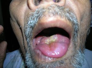
Tongue should be carefully examined. The patient's ability to protrude the tongue is also ascertained. If the patient has tongue tie then full protrusion of tongue is not possible. Size of the tongue must also be seen. Macroglossia is seen in acromegaly / Down's syndrome.
In hypoglossal nerve palsy the tongue deviates to the side of the lesion. The tongue on the paralysed side may show wasting of lingual musculature. Fasciculation of tongue is seen in motor neuron disease.
Loss of papilla is seen in patient's with vitamin deficiency, in those patients who have under gone irradiation of that area.
Malignancy of tongue is common over its lateral surface. Any suspicious swelling of tongue must be palpated for signs of induration, which is a characteristic feature of malignant lesions of tongue.
Tongue coating is seen in cases of oral thrush and in patients with febrile illness.
Opening of the wharton's duct can be seen under the tongue. If there is swelling in this area then it must be palpated to rule out submandibular gland calculus.
The opening of the parotid duct can be examined after gently retracting the cheek. It lies opposite to the upper second molar.
Palate is examined for ulcers, clefts, perforations or presence of masses.
The position of the uvula must be seen. Normally uvula is in the midline. In cases of palatal paralysis uvula deviates to the opposite side.
Tonsillar pillars must be clearly seen. It is commonly congested in chronic tonsillar infections. Tonsils must be examined. Its size must be noticed. Tonsillar enlargement can be classified under the following heads:
Grade 0 - Tonsils are found confined to the space between the anterior and posterior pillars
Grade 1 - Tonsils are enlarged and is just seen coming out of the anterior pillar.
Grade 2 - The enlarged tonsil reaches to about half the distance of uvula.
Grade 3 - The enlarged tonsil comes into contact with the uvula.
Grade 4 - The enlargement of tonsil is so much that both tonsils lie virtually in contact with each other i.e. kissing tonsils.
Hypopharynx include:
1. Posterior pharyngeal wall
2. Pyriform fossa
3. Post cricoid region
Examination of this area is done by
1. Indirect laryngoscopy
2. Flexible and rigid endoscopy
3. Imaging
Indirect laryngoscopy:
1. The mirror used is plane mirror with a long handle.
2. It is held like a pen in the dominant hand with the mirror pointing downwards.
3. The mirror is warmed with a spirit lamp, the temperature is tested on the back of the hand
4. The patient is asked to protrude the tongue and it is held with a gauze.
5. The mirror is introduced into the mouth and gently slide under the uvula.
6. The mirror is tilted to get good view of the larynx.
7. The patient is asked to say eee.
8. The mobility of the vocal cord can be tested.


