Childhood stridor
Stridor is defined as noisy breathing.
Causes of childhood stridor:
Causes of stridor are anatomically classified:
1. Supralaryngeal causes:
a. Nose - choanal atresia
Obstruction due to infection / truama / tubes
b. Cranio facial anamolies:
These patients have narrowing of oropharynx, nasopharynx andnasal cavities. The may also additionally manifest with macroglossia.The various anamolies associated with respiratory difficulties are:
Pierre Robin syndrome
Treacher collin syndrome
Apert's syndrome
Cruzon's syndrome
Mobieus syndrome
c. Macroglossia :
Beckwith Wiedemann syndrome
Down's syndrome
d. Tumors:
Hemangioma
Neuroblastoma
e. Laryngomalacia : Is caused by an excessiely elastic cartilagenous support to the airway seen in infants. This commonly affects the glottic and supra glottic airway of infants. This excessively soft and elastic cartilage causes inspiratory collapse ofthe arytenoid, aryepiglottic folds and epiglottis during inspiration.The omega shaped epiglottis seen often in the infants adds to theproblem. This causes occlusion of the laryngeal inlet. These patient shave inspiratory stridor which becomes better on prone position or when the child is calm. Stridor is worsened if the child is restless orexcited. The cry of the child is usually normal. The child may also have aspiration and feeding difficulties. It is commonly seen during the first few months of life.
2. Glottic causes:
a. Vocal cord palsy : Is one of the commonest cause of airway obstruction. In 80% of patients it is unilateral.
Etiology: Could be caused due to injury to vagus nerve at the level of Nucleus ambiguus - it is often bilateral.
Injury to the left recurrent laryngeal nerve due to cardio vascular causes and thoracic causes. It could be caused due to increased intracranial pressure - i.e. Meningomyelocoele with Arnold Chiari malformation.
Clinical features: Inspiratory stridor at birth Weak, hoarse cry or aphonia.
If unilateral the patient feel better when placed on the side of the lesion.
b. Tumors :
Papilloma
Hemangioma
Cystic hygroma
Laryngoceles
c. Atresia
d. Webs: are caused due to failure of recanalisation of thelarynx . It can range between a complete occlusion by mucosa andsubmucous tissue or partial occlusion by a thin membranous web. It canoccur in supraglottis, glottis and subglottis area. Commonly it is seenin the glottic area. It occurs in one in 10,000 live births.
Indications of tracheostomy:
"The main indication of tracheostomy is that when the surgeon thinks about it" (Mosher).
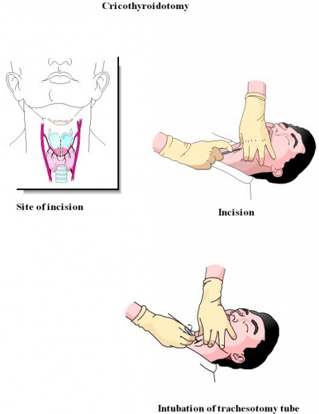
1. In upper air way obstruction (obstructionabove the level of larynx). Trachesotomy is indicated in all cases ofupper airway obstruction irrespective of the cause as an emergency lifesaving procedure. It is also indicated in impending upper airway obstruction as in the case of angioneurotic oedema of larynx.
2. For assisted ventilation: In comatose patients who donot have the required respiratory drive airway can be secured by performing a tracheostomy and the patient can be connected to a ventilator for assisted ventilation.
3. For bronchial toileting: Chronically ill patients who donot have sufficient energy to cough out the bronchial secretions may have to undergo tracheostomy with the primary aim of sucking out the bronchial secretions through the tracheostome.
4. In cases of prolonged intubation: tracheostomy will have to be performed to prevent subglottic stenosis. Complications of tracheostomy:
1. Injury to thryoid isthumus causing troublesome bleeding
2. Too lateral dissection may cause extensive bleeding and possible injury to recurrent laryngeal nerve.
3. Injury to the apex of the lung (right)
4. Sudden apnoea when the trachea is opened,due to loss of hypoxic respiratory drive. This can be prevented by slow opening of the trachea, or by subjecting the patient to inhage carbogena mixture of carbondioxide and oxygen.
5. Subcutaneous emphysema if pretracheal fascialis not dissected properly, or too small a tube is introduced into the tracheostome.
6. Injury to great vessels. This can occur in children.
