Branchial anamolies
Contents
Definition:
Branchial apparatus develop between the 3rd and 7th weeks of embryonic life. These structures are phylogenetically related to the gill slits of fish. To begin with there are 5 mesodermal arches separated by invaginations of ectoderm (clefts) and endoderm (pouches). Each of these archeshas its own unique arterial and nervous supply. These structures eventually develop into muscles and connective tissue structures of neck.
The components of branchial apparatus include:
1. Branchial arches
2. Branchial clefts
3. Branchial pouches
4. Their blood and nerve supplies
l: Lateral tubercle, IM: tuberculum impar, c: foramen cecum, T: thyroglossal duct, e: cervical sinus. The cartilages, nerves and blood vessels are marked accordingly
Branchial arches:
The branchial arches are 6 in number. Each of these arches are made up of a core of mesodermal tissue covered on the outside by surface ectoderm, and on the inside by endoderm. Each of these arches has its own cartilagenous bar, muscular component, arterial, venous and nerve supplies. Each arch in addition to its nerve supply also receive branches from the nerve supplying the succeeding arch. Hence each arch receives a branch , called the post-trematic from the nerve of its own arch, and a second branch called the pre-trematic from the succeeding arch.
During development the second arch grows caudally to cover the third and fourth arches and the second, third, and fourth pharyngeal clefts eventually fusing with the lower neck. The enclosed II, III, and IV clefts are called as the cervical sinus. The buried clefts (cervical sinus) persist as cavities lined by ectoderm and gradually disappears with development. If this process does not occur for some reason then it gives rise to branchial cyst, sinus or fistula.
| Arch | Post-trematic nerve | Pre-trematic nerve |
|---|---|---|
| First | Mandibular nerve | Chorda tympanic branch of facial nerve |
| Second | Facial nerve | Tympanic branch of glossopharyngeal nerve |
| Third | Glossopharyngeal | Pretrematic neves not well defined |
| 4th, 5th | Vagus and accessory nerves | |
| 6th | Vagus and accessory nerves |
Branchial anamolies:
Include branchial cysts, sinuses, and fistulae. It is now considered that these anamolies are not variants of the same anamoly involving the branchial apparatus, but are entirely different in their pathogenesis.
Branchial cysts:
also known as lateral cervical cysts usually present in the lateral portion of the neck deep to the sternomastoid at the junction of its upper 1/3 and lower 2/3. Some of these cysts may have a tract up to th posterior pillar of tonsil. These cysts usually smooth, round, non tender fluctuant mass. Males and females are equally affected. Secondary enlargement of these cysts are common during upper respiratory infections due to the enlargement of lymphopid tissue found lining the cyst wall. Patients usually present between the second and fourth decades of life. Depending on its size it could produce dyspnoea, dysphonia, dysphagia and cosmetic deformity.
To summerize:
Clinical features:
1. Continuous swelling
2. Intermittent swelling
3. Pain
4. Infection
5. Secondary infections
In rare circumstances abscess may occur in a branchial cyst leading to rupture of the contents and sinus formation.
Histology:These cysts are lined by respiratory or squamous epithelium. During episodes of infections inflammatory cells can be commonly seen. Lymphoid tissue may also be present beneath the lining epithelium.
Differentiatial diagnosis:
Branchial cysts should be differentiated from cystic hygroma, lymph cyst, carotid body tumors, ectopic salivary glands, neurofibrama etc.
Theories of orgin of branchial cyst:
There are four known theories of origin of branchial cysts. Due to the complicated nature of the embryology of the neck none of these theories have been proven to be right.
1. Branchial apparatus theory:
According to this theory, these cysts may represent the remains of the pharyngeal pouches or branchial clefts or a fusion of these two elements. When these cysts have an internal opening, it lies in the posterior pillar of the tonsillar fossa thus attributing its origin to the second branchial pouch. Fistulae and sinuses from the second pouch would necessarily pass between the external and internal carotid arteries.
An origin from the third or fourth pouches is unlikely since they have to pass over the hypoglossal nerve to reach the skin and would be severed by the upward movement of that nerve during development. A third arch tract should have its internal opening at the level of pyriform fossa, while the fourth arch tract will have to open below this level. These openings have never been described so far. The fourth tract would have to pass below the subclavian artery on the right and and aortic arch on the left side. Origin from these pouches has been discounted.
Origin from the first pouch is a distinct possibility, high branchial cysts have been described lying under the parotid gland with an internal opening between the cartilaginous and bony external auditory canal. The peak age incidence between the third and fourth decades is pretty late for a congenital lesion.
2. Cervical sinus theory:
This theory postulates that branchial cysts represent the remains of cervical sinus of His which is formed by the second arch growing down to meet the fifth arch. If this theory is true then internal opening for these cysts is not possible.
3. Thymopharyngeal duct theory:
This theory postulates that branchial cyts represent remains of original connection between the thymus and the third branchial pouch. This theory also assumes that the hyoid bone constituted the lower level of branchial derivatives. This is ofcourse false, and a persistant thymic duct has never been described. No branchial cysts has ever been described deep to the thyoid gland making this theory unacceptable.
4. Inclusion theory:
was described by King. He suggested that the cyst developed from the branchial apparatus and the cyst epithelium arose from lymph node epithelium. The following facts lend credence to this theory:
a. Most branchial cysts have lymphoid tissue in their wall and are found in th parotid, pharynx as well as lateral neck.
b. The peak age incidence is later than expected for a congenital lesion.
c. Most branchial cysts have no internal opening, or at best a tract with ill defined termination.
Histology:
The branchial sinus and fistula are lined by stratified squamous epithelium, occassionally they could be lined by non ciliated columnar epithelium. If it is lined by non ciliated columnar epthelium, it can be safely assumed to be glandular metaplasia due to infection. This glandular tissue starts to secrete mucinous material filling up the cyst cavity. Some of the cysts contain straw colored fluid, which could have come only from the blood (transudate).
Management:
These masses if present should be removed surgically for:
1. Confirming the diagnosis
2. Cosmetic reasons
3. To prevent infections
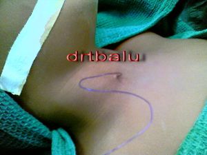
Surgical removal is performed using a transverse skin crease incision. The sternomastoid muscle is retracted. The cyst is mobilized and an attempt is made to identify the tract. Before surgery a careful examination should be performed to identify inner opening close to the posterior pillars of the tonsil.
Branchial sinus / fistula:
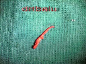
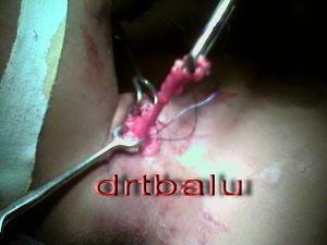
The mouth of the sinus should be encompassed in the incision, preferably elliptical. The tract if present will be sometimes as thick as a big artery. If possible effort sould be made to canalize the fistula using proline, this will help in totally identifying the complete extent of the tract. The tract could pass between the internal carotid artery and internal jugular vein. It should be completely excised to prevent recurrence.


