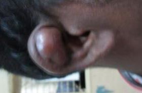
Auricle Trauma
Introduction:
Injury to auricle has been documented since Roman times when judicial removal of the auricle for theft was practised to deter thieves. Self mutilation of pinna for insertion of ear ring has also been documented. Ear piercing indeed carries the risk of perichondritis and spread of viral diseases like hepatitis and AIDS.
Auricle has a complicated shape because of the presence of irregular plate of elastic cartilage which is about 0.5 - 2 mm thick. The skin covering of the auricle has a subcutaneous layer only on its posterior medial convex surface. It displays few hairs which are infact small. Sebaceous glands can sometimes be of considerable size by sweat glands are scarce. The lobule of the ear does not have a cartilage framework but contains only fat tissue.
Accidental injury of the auricle are known to occur in all age groups, but persons participating in contact sports like wrestling, boxing and foot ball are more prone to pinna injuries. Pinna injury in a child should always raise the question of child abuse.
Trauma to auricle may result in laceration / partial / complete loss.
Spectrum of auricular injuries:
Injury without tissue loss:
Hematoma of the auricle is the result of closed trauma to pinna. This occurs commonly in persons engaged in contact sports. This results in disfigurement by fibrosis / chondritis.
Injury with tissue loss:
External injury to pinna may be associated with tissue loss/uncomplicated rupture / perforation of ear drum / fracture of the petrous portion of temporal bone.
Aural seroma:
This is an acquired condition involving the pinna due to trauma. Blunt trauma is commonly known to cause this problem. Wrestlers and boxers are commonly affected. It can also occur without any cause (idiopathic). Intense allergic reaction can also cause aural seroma
Aural seruma is caused due to collection of blood (hematoma auris) / serous fluid between the aural cartilage and perichondrium stripping it away from the cartilage compromising its nutrition. Untreated aural seroma can ultimately lead to deformities like cauliflower ear. It is also known as pseudo cyst of the pinna. This term was coined by Hartmann in 1846. Commonly it contains serous fluid.
Symptoms:
1. Heaviness in the pinna region
2. Swelling in the pinna region
Cystic swelling is commonly located in scaphoid fossa of pinna. It is common in males than in females. It has been postulated that pseudocyst formation could be due to disruption of aural cartilage.
Management:
Ideal management modality is incision and drainage. It is however prone for recurrence. The fluid collected between the cartilage and perichondrium is usually sterile.
Small lesions can be aspirated using syringe. After aspiration compression dressing is applied sometimes even plaster of paris is preferred.
Window operation:
This surgical procedure peformed in recurrent aural seromas. A window is created over the most prominent region of the swelling and teh fluid is drained. Compression dressing is applied.
Perichondritis is the common complication of surgical procedure involving the pinna. Patient should be placed under antibiotic cover following surgery.
Pathophysiology current concepts:
Serous fluid effuion originates from the cartilage. In the early stages of the lesion the top wall of the cyst is formed by the cartilage membrane. This membrane keeps proliferating as the size of the cyts increases. New cartilage formation occurs from this membrane. Eventually, the secretion is walled on all sides by the cartilage. Effusion during this stage becomes intracartilaginous. Finally the fluid gets absorbed and the new cartilage gets adherent to the cartilage pinna. The auricle eventually becomes thickened and deformed due to this process.
Three phases have been identified in pseudocyst of pinna. They are:
Early period - Acute exudative period
Medium period - Cartilage formation period
Late period - proliferative and organized period
Local injections of steroids have been proven to be effective and more and more surgeons are preferring to use steroid infiltration as a first step in the management of aural seroma.
Simple lacerated injury of pinna:
Can be closed under aseptic conditions using either skin to skin vicryl sutures or if needed skin sutures combined with inter cartilaginous sutures using fine suture materials. Unnessary suturing of pinna cartilage is to be avoided as it could provide a pathway for bacterial infection.
Cases of meatal stenosis were succcessfully treated with split thickness graft.
Totally torn ear should accurately be replaced within 2 hours of injury. If cartilage is bare and with intact perichondrium skin graft is essential. It is very important to prevent drying of the exposed cartilage by use of topical antibiotic. One exception to primary closure is when the ear has been injured by human bite.
