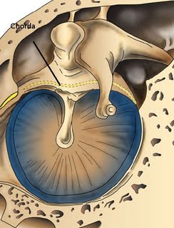
Anatomy of chorda tympani nerve
Introduction:
The chorda tympani nerve is a branch of facial nerve. It exits the facial nerve just before it exits via the stylomastoid foramen. It is one of the three cranial nerves that is involved in transmission of taste fibers from the tongue. Chorda tympani nerve conveys taste fibers from the anterior 2/3 of tongue. Mechanism of taste sensation is rather unique in that it involves a complicated feed back loop with each nerve acting to inhibit the signal of other nerves. The chorda tympani exerts strong inhibitory influence on other taste fibers as well as pain fibers from the tongue. When chorda tympani nerve is damaged its inhibitory function is disrupted, causing the other taste fibers to act in an uninhibited manner.The chorda tympani nerve carries with it two types of fibres which traverse via lingual nerve to reach their destination. These fibers include:Special sensory fibers providing taste sensation from anterior 2/3 of tongue.
Presynaptic parasympathetic fibers to submandibular ganglion providing secretomotor fibers to submandibular and sublingual salivary glands
Presynaptic parasympathetic fibers is also supplies the blood vessels of the tongue. When stimulated the chorda tympani nerve causes dilatation of blood vessels of the tongue.
Central connections:
Chorda tympani nerve contains fibers from two brain stem nuclei. They are:
Superior salivatory nucleus:
This nucleus houses cell bodies of secretomotor preganglionic parasympathetic neurons
Nucleus of tractus solitarius:
The superior portion contributes to chorda tympami fibers. It receives central processes of taste neurons which have their cell bodies in the ganglia of the three cranial nerves conveying taste sensation. After synapsing in this nucleus secondary axons ascend in the lateral lemniscus to relay in the thalamus.
This pathway then passes through the posterior limb of internal capsule to reach the primary gustatory cortex.
Connections seen within the facial canal:
Sensory branch of facial nerve Nervus intermedius of Wrisberg joins the facial nerve here. It conveys special sensory fibers from taste buds present in the anterior 2/3 of tongue and soft palate. It also contains secretomotor fibers to salivary glands present below the oral cavity. Nerves intermedius exits the brain stem adherent to the vestibuo cochlear nerve. At the level of internal auditory meatus it leaves this nerve to merge with that of facial nerve.
Chorda tympani nerve exits from the facial nerve before the facial nerve exits via the stylomastoid foramen. It is the largest branch of facial nerve in its intrapetrous compartment. It arises below the nerve to stapedius. It traverses antero superiorly via the posterior canaliculus usually accompanied by posterior tympanic branch of stylomastoid artery. This canaliculus opens into the middle ear cavity through an aperture situated at the junction of posterior and lateral walls of tympanic cavity. This opening lies just medial to the fibrocartilagenous annulus. The posterior canaliculus is roughly 0.5 mm in diameter. Chorda tympani nerve shows a large number of variations. In some patients the chorda tympani nerve may arise from more proximal portion of facial nerve, even close to the geniculate ganglion. The length of the posterior canaliculus is also highly variable ranging from 3 – 14 mm. In 10% of individuals there may not be a posterior canaliculus at all but could be replaced by a groove.
If the chorda tympani nerve originates outside the temporal bone then the posterior canaliculus will be separate from that of the facial nerve canal. In fetus and young infants the chorda tympani nerve leaves the facial nerve outside the skull, but the postnatal growth of mastoid process causes it to migrate to a more proximal position. Since the facial canal grows more than the mastoid segment of facial nerve the chorda tympani nerve typically diverges from the facial nerve in an infant of 1 yr of age.
Course of chorda tympani in the tympanum:The chorda tympani arches across pars flaccida medial to the upper part of the handle of malleus and traverses above the insertion of tensor tympani. In patients with congenital anamolies of malleus chorda is also displaced laterally.Chorda tympani nerve exits the middle ear via a separate bony canal, the anterior canaliculus also known as the canal of Hugier. This canal runs in the medial portion of petrotympanic fissure. Anterior tympanic branch of maxillary artery accompanies this nerve along this canal. Chorda exits the skull through a small foramen behind the base of spine of the sphenoid. At its exit it is closely related to the medial surface of temporomandibular joint.
Course of Chorda in the infratemporal fossa:
In the infratemporal fossa the chorda tympani nerve descends medial to the spine of sphenoid and angles forward to join the lingual nerve about 2 cms below the skull base.
This junction lies close to the lower border of lateral pterygoid muscle.
Functions of chorda tympani nerve:
1. It carries taste sensation from the anterior 2/3 of tongue
2. Supplies secretomotor fibers to the salivary glands in the floor of the mouth
3. Conveys general sensation from the anterior 2/3 of tongue which includes pain and temperature Supplies secretomotor fibers to the parotid gland
4. Supplies efferent vasodilator fibers to the tongue
