Anatomical changes that occur in ethmoid sinuses following FESS
Introduction:
The bony walls of paranasal sinuses demonstrate excellent degree of plasticity. This feature allows alterations in the size and dimensions of paranasal sinuses during growth phase. The process of pneumatization of paranasal sinuses begins in utero and continues through teenage years.
Causes of pathological expansile alterations of paranasal sinuses include:
Allergic fungal sinusitis
Extensive sinonasal polyposis
Mucocele formation
Benign tumors
Pathological causes of contractile changes in sinus cavity:
Cystic fibrosis
Silent sinus syndrome – caused due to obstruction of sinus ostium leading on to atelectasis of maxillary sinuses.
While performing endoscopic sinus surgery it should be borne in mind that it is being performed in a setting of bony changes, with an intention to halt the expansile / contractile changes that are likely to take place.
While performing endoscopic sinus surgery in paediatric age group it should always be borne in mind that there is a real time risk in causing irreversible bony changes in a growing bone causing undesired changes in the facial morphology. Recent studies however counter this claim saying that it is very safe to perform ESS in peadiatric age group.
Anatomical changes that take place in the ethmoidal sinuses of adult patients who have undergone ESS:
This is a largely understudied topic. Few studies that have been performed by analysing the axial CT scans pre op and post op has demonstrated that there is bowing of lamina papyracea following surgery. This medial bowing caused a reduction in the volume of ethmoidal sinuses by 1mm cube. The amount of bowing was proportional to the extent of surgery. Maximum changes in the ethmoid sinus dimensions occured at the level of posterior globe.
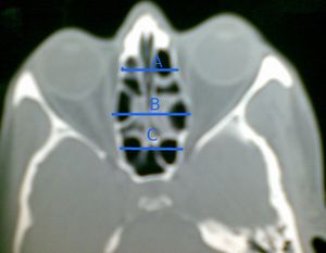
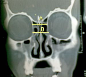
Michel Platt etal in their work have suggested that a total of 5 measurements should be made before coming to a conclusion that there is a definite change in the dimensions of ethmoidal sinuses. These measurements should be made in the pre op and post op CT scans of the patients.
The distance between both lamina papyracea were measured in the axial plane at the level of planum sphenoidale.
The distance between both lamina papyracea are measured in the axial plane at the mid globe level.
The distance between both lamina papyracea are measured in the axial plane at the posterior globe level
The distance between both lamina papyracea are measured at the level of anterior wall of sphenoid sinus
In the coronal plane the authors advised measurements to be made consistently at the:
Plane of posterior globes
Level of cribriform plate
At the length of 5-10 mm below the cribriform plate

