An incidental case of bilateral choanal atresia
Contents
Patient history:
18 years old male
Bilateral nasal obstruction since childhood ++
Right sided nasal discharge since childhood ++
Clinical examination:
Deviated septum to left ++
Right inferior turbinate hypertrophy ++
Imaging:
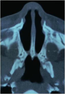
CT scan PNS plain coronal cuts showed:
DNS
Rt inferior turbinate hypertrophy
There was no evidence of sinusitis
Choanal atresia could not be made out ? missed out in the cuts i.e. 5 mm cuts
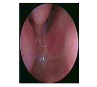
DNE showed:
DSL with inferior turbinate hypertrophy
Choanal atresia on both sides
Treatment:
Septal correction with transnasal repair of choanal atresia was performed Atretic bony plate was drilled out and the recreated choana was stented.
Post op management:
Stent was left in place for 6 weeks.
Regular nasal douching and suction clearance was done.
Discussion:
During the third week of IU life the nasal placodes starts to invaginate forming nasal pits.
These pits burrow into the surrounding mesenchyme forming nasal pouches.
Formation of bucconasal membrane occurs at this point.
Pathogenesis:
Choanal atresia could be caused by persistent bucconasal membrane / faulty mesodermal migration.
Boundaries of atretic plate:
Superior - sphenoid
Lateral - Medial pterygoid lamina
Medial - vomer
Inferior - Horizontal plate of palatine bone
CT scan features suggestive of choanal atresia:
Choanal air space measurement:
Mean normal - 0.67
Mixed atresia - 1/3 of normal
Bony atresia - 0
Mean width of vomer:
Mean - 0.23
Bony atresia - 0.6
Membranous atresia -0.3
Possible surgical approaches:
Transnasal
Transpalatal
Trans septal
Syndromes associated with choanal atresia:
CHARGE
Treacher Collins
Crouzans

