Surgical Management of Glomus jugulare
Introduction:
Glomus tumors of temporal bone occur in the region of the jugular bulb and middle ear. These are very rare, vascular, slow growing tumors. They are most often benign. Tumors originating from the jugular bulb and extending to involve the middle ear cavity are referred to as glomus jugulare tumors.
These tumors are more common in females i.e. thrice as common than in males. It is more common on the left side. Most of these tumors occur in patients in the age group between 40-70 years.
Etiology:
These tumors originate from the chief cells of the paraganglia / glomus bodies located within the wall (adventitia) of the jugular bulb. These lesions can also be associated with the auricular branch of vagus nerve (Arnold's nerve) or the tympanic branch of the glossopharyngeal nerve (Jaconson's nerve).
Classification:
Glasscock-Jackson and Fisch classification is widely used in glomus tumors. This classification is based on extension of the tumor to the surrounding anatomic structures and is closely related to mortality and morbidity.
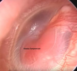
Type A tumor - Tumor limited to middle ear cleft (glomus tympanicum)
Type B tumor - Tumor limited to the tympanomastoid area with no infralabyrinthine compartment involvement
Type C tumor - Tumor involving the infralabyrinthine compartment of the temporal bone and extending into the petrous apex
Type C1 tumor - Tumor with limited involvment of the vertical portion of the carotid canal
Type C2 tumor - Tumor invading the vertical portion of the carotid canal
Type C3 tumor - Tumor invading the horizontal portion of the carotid canal
Type D1 tumor - Tumor with intracranial extension of less than 2 cm in diameter
Type D2 tumor - Tumor with an intracranial extension greater than 2 cm in diameter
Surgical management of glomus jugulare:
Surgery happens to be the treatment of choice for glomus jugulare tumors. Radiation therapy, particularly stereotactic radiosurgery is showing more promise because of low risk of treatment related cranial nerve injury. In managing large lesions surgically pre operative embolism could make the job of the surgeon a little easier.
Type A tumors:
Also known as glomus tympanicum because the mass is confined to the middle ear cavity. Transcanal resection of the mass is the treatment of choice in these patients.
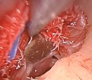
The surgery is ideally performed under general anesthesia with hypotension. Tympanomeatal flap is elevated as in the case of middle ear surgeries. The mass would become visible once the middle ear is entered. The mass can be removed completely after cauterization of its vascular supply. The tympanomeatal flap should be competely detached from the handle of malleus to facilitate better visualization of the tumor mass.
Conservative jugulopetrosectomy:
This approach preserves the normal anatomy of external and middle ear. It is indicated for small tumors confined to the jugular foramen and infralabyrinthine area with minimal involvement of internal carotid artery.
Skin incision is placed about 6 cm behind the postauricular crease and the same incision is extended inferiorly and superiorly to expose the neck and temporal bone. The 7th,9th, 10th, 11th and 12th cranial nerves are followed up to the skull base. The facial nerve is dissected until its bifurcation. Major neck vessels are identitied. The internal jugular vein is ligated and divided to obtain distal control of the vein. A complete enlarged mastoidectomy is performed and the posterior wall of the bony ear canal is preserved. Lateral sinus is exposed from the transverse sinus to the jugular bulb and then opened and packed intraluminally with surgicel to obtain proximal control of the lateral sinus.
The facial nerve is identified and skeletonized to the stylomastoid foramen. An extended facial recess approach is drilled to enable the initial visualization of the tumor. When the external canal is preserved, the facial nerve is mobilized from the second genu laterally to expose the jugular bulb. Further inferior widening of the extended facial recess and removal of tympanic bone anteroinferiorly exposes the lower part of the tympanic segment of the internal carotid artery (infratubal portion) and the carotid foramen. Once the internal carotid artery is freed from the tumor, it can be mobilized from the infralabyrinthine area. With the internal jugular vein and the lateral sinus identified, the tumor is resected from the skull base and cranial nerves. If possible the medial wall of the jugular bulb is left intact to avoid danger to the cranial nerves.
Infratemporal fossa approach:
This approach is indicated when the mass extends beyond the jugular foramen anteriorly into the petrous bone. This approach is indicated if the distal exposure of petrous carotid artery is needed. This approach sacrifices the external and middle ear. It offers access to the infralabyrinthine area, petrous apex, and through the infratemporal fossa, to the entire petrous carotid artery, clivus and cavernous sinus.
1. Preparation - Head should be shaven free from hair.
Facial nerve monitoring
Patient positioning - supine with head elevated and face turned towards the opposite side.
Draping
2. Ear canal resection
3. Mastoidectomy
4. Jugular vein ligation
5. Facial nerve exposure
6. Exposure of jugular bulb
7. Intracranial dissection
8. Closure
The petro-occipital skull base, neck, and infratemporal fossa are accessed through a C shaped incision that allows an anteriorly based flap. The external ear canal is then transected the sutured. Both temporozygomatic and cervicofacial branches of the facial nerve are exposed to allow sufficient mobilization of the nerve from the parotid for anterosuperior transposition. The skin of the external auditory canal, ear drum and the ossicles are removed. A radical mastoidectomy is performed. The lateral sinus, middle fossa dura, and facial nerve are identified.
Lateral sinus is unroofed. The internal jugular vein is exposed and ligated in the neck. Lateral sinus is opened and packed intraluminally. Exposure of the petrous portion of internal carotid artery requires anterior dislocation of the ascending ramus of the mandible, this also requires anterosuperior rerouting of the facial nerve. This maneuver is a must to obtain satisfactory control of jugular foramen and petrous carotid artery. This allows exposure of the glenoid and posterior infratemporal fossae. Removal of the medial wall of the glenoid fossa and of the bony eustachean tube exposes the vertical portion of petrous carotid artery and the anterior pole of the tumor. Subtotal petrosectomy permits control of the superior pole of the tumor and its radical dissection from the cancellous bone of the petrous apex and from the petrous carotid wall.


