Difference between revisions of "Sphenoidal fungal sinusitis with middle cranial fossa extension"
From Otolaryngology Online
(Created page with " Category:Case Report Case History: 45 years old female patient was admitted with C/O: 1. Altered consciousness - 2 days duration History elicited: 1. Headache inte...") |
|||
| Line 36: | Line 36: | ||
Debridement was done endoscopically under general anesthesia. | Debridement was done endoscopically under general anesthesia. | ||
Patient had complete recovery following surgery. | Patient had complete recovery following surgery. | ||
| − | |||
| − | |||
Latest revision as of 10:47, 15 May 2019
Case History:
45 years old female patient was admitted with
C/O:
1. Altered consciousness - 2 days duration
History elicited:
1. Headache intense more in the occipital area - 2 months
2.Low grade fever - 20 days (for which the patient received intermittent treatment)
3. Patient had vomiting - 3days back
Patient is a known diabetic on regular treatment with oral hypoglycemics
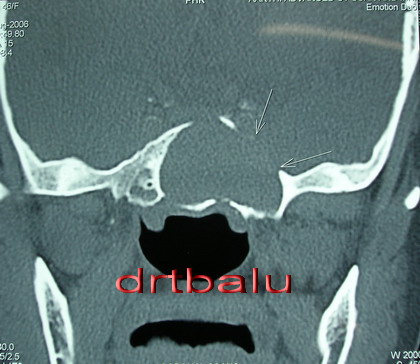
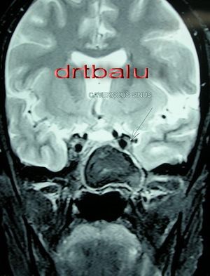
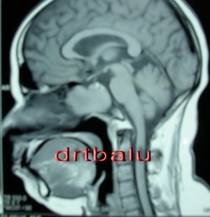
Imaging:
Management:
Debridement was done endoscopically under general anesthesia. Patient had complete recovery following surgery.


