Difference between revisions of "Interesting case of fibroma sphenoid sinus"
(Created page with " Category:Case Report === Clinical details: === 18 years old boy came with c/o blocking sensation both nasal cavity - 1 year duration Decreased sensation of smell - 6...") |
|||
| Line 38: | Line 38: | ||
Fibroma involving sphenoid sinus and post nasal space are commonly vascular (angiofibroma). In this case the fibrous elements predominated and hence the mass was not enhancing in contrast CT. There was no abnormal bleeding during surgery. | Fibroma involving sphenoid sinus and post nasal space are commonly vascular (angiofibroma). In this case the fibrous elements predominated and hence the mass was not enhancing in contrast CT. There was no abnormal bleeding during surgery. | ||
| − | |||
| − | |||
| − | |||
| − | |||
| − | |||
| − | |||
| − | |||
| − | |||
Latest revision as of 10:43, 15 May 2019
Clinical details:
18 years old boy came with c/o blocking sensation both nasal cavity - 1 year duration
Decreased sensation of smell - 6 months duration
He gave h/o repeated attacks of upper respiratory infection
He gave no h/o bleeding from nose
No h/o head ache
On examination:
Anterior rhinoscopy:
A pale mass could be seen occupying the posterior portion of right nasal cavity.
Post nasal examination:
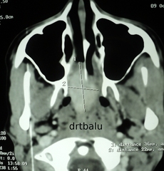
The same mass could be seen occupying the nasopharynx occluding both choana. CT scan nose and paranasal sinuses plain / contrast were taken.
It showed non enhancing mass arising from sphenoid sinus entering the post nasal space and the posterior portion of right nasal cavity
Management:
This patient was taken up for endoscopic sinus surgery and the mass was removed completely in toto.
Histopathology report - fibroma
Fibroma involving sphenoid sinus and post nasal space are commonly vascular (angiofibroma). In this case the fibrous elements predominated and hence the mass was not enhancing in contrast CT. There was no abnormal bleeding during surgery.
